Download
1 / 54
550 likes | 747 Vues
Sensory and Motor Systems. Sensory System Principles. Specialized receptors Limited receptive fields Localization of stimuli Coding/Preprocessing Complex neural pathways Multiple representation in cortex. Sensory System Principles. Specialized receptors

E N D
Sensory System Principles • Specialized receptors • Limited receptive fields • Localization of stimuli • Coding/Preprocessing • Complex neural pathways • Multiple representation in cortex
Sensory System Principles • Specialized receptors • Neurotransmitter receptors are replaced with chemical- or mechanical motion- sensitive ion gates which cause neuron depolarization, and in most cases, action potentials. • Two general types • Slow adapting • Sense stimulus for a longer period. • Fast adapting • Sense stimulus changes for a short period.
Sensory System Principles • Limited receptive fields • Each sensor responds to only part of the world. • Localization of stimuli • Localization by comparison of different views. • The wider the sensor separation, the better the localization.
Sensory System Principles • Coding/Preprocessing • Very little sensory information is used by the nervous system exactly as it is transduced. • High compression: many sensors, few neurons. • Preprocessing and feature extraction. • Allows the cortex to concentrate on recognition, planning and response.
Sensory System Principles • Complex neural pathways • Most neuromuscular pathways are 1-2 neurons • Most sensory pathways are 3-4 neurons • More responses on different levels. • Sensory systems can drive multiple centers. • Multiple representations in cortex • The same sense can be represented in multiple ways in the cortex. • Often related to the sensory feature extraction. • Separate cortical areas for each sense.
The Senses • Vision (sight) • Olfaction (smell) • Gustation (taste) • Audition (hearing) / Vestibular (orientation) • Touch • Proprioception (body position) • Nociception (pain) • Chemosensors (BP, glucose, acidity, etc.)
Vision • Probably one of the most important senses. • Vision occupies a greater percentage of the human cerebral cortex than any other sense. • Almost all organisms are light sensitive and can react to light intensity and/or direction. • Ancient sense, possibly derived from a symbiotic bacterial inclusion.
Vision • Limited spectral range • Humans see electro-magnetic radiation in the 380-760 nm range, which corresponds to peaks of solar radiation. • Insects can see UV. • Snakes can see IR (heat). • Polarized vision in some species.
Vision • Eye anatomy • Protective layers • Cornea & sclera • Deformable lens • Fluid filled • Photosensitive retina, optic nerve and blood supply in rear
Vision • Retina • Rods • Hi-sensitivity BW vision • Cones • Low-sensitivity color vision • Blood supply • Several layers of processing neurons • Light-sensitive cells involved in circadian rhythms, not vision • Tapetum in some species
Vision • Rods • Night vision • Slowly adapting • 120 million • Not in fovea • Cylindrical outer segment with stack of disks w/receptors • Only one opsin • Rhodopsin 498 nm (3)
Vision • Cones • Color vision • Fast adapting • 6 million, mostly in fovea • Conical outer segment of stacked disks • Three opsins • Cyanolabe 437 nm (7) • Chlorolabe 533 nm (X) • Erythrolabe 564 nm (X)
Vision • Vision receptors • 7 TM receptor with retinal-opsin sensory complex. • Retinal is derived from Vitamin A. • Opsin is frequency (color) sensitive. • Light stimulation changes retinal conformation & 2nd messenger system. • Releases glutamate
2nd messenger transduction • Single photon can be detected! • Rods and cells produce graded potentials, no APs!
Presence of cGMP opens Na+ channels Dark cGMP keeps Na+ channels open Cell is depolarized Light 2nd messenger system turns cGMP into GMP No cGMP, Na+ channels close Cell hyperpolarizes
Vision • Circadian sensor cells • Newly discovered light-sensitive cells in the inner ganglion layer of the retina project directly to the suprachiasmic nucleus (SCN), site of the human circadian clock. • Sensitive pigment is melanopsin - a newly discovered opsin (chromophore, derived from vitamin B2).
Vision • Retinal processing • Receptors • Graded potentials, gap junctions, hyperpolarize in light • Horizontal cells • L-type (luminosity) hyperpolarize in all light • C-type (color) hyperpolarizes only for certain colors • Only inhibitory • Bipolar cells • Concentric oppositional surround response • 1:1 bypass for foveal receptors only
Vision • Retinal processing • Amacrine cells • Seem to be responsive to changes • Gets input from midbrain in some species • Ganglion cells • First to generate action potentials • 800,000 outputs (150:1 for rods, 8:1 for cones) • X (slow moving objects in fovea) • Y (fast moving objects in periphery) • W (orientation specific, vestigal in humans) • M (moving object detection, transient) • P (form, color, and fine detail, sustained)
- + - - • + + + - • - + - -
Vision • Anopsias • Partial field of view • Hemianopsia = ½ field • Lesions before LGN • Semi-hemianopsia = ¼ of field • Lesions after LGN 1 3 2 4, 5
Vision • Lateral Geniculate Nucleus (LGN) in thalamus: • 80% of optic nerve • Left hemi-fields of both eyes project to right LGN of the thalamus and then onto the V1 cortex.
Both the LGN and Superior Colliculus are mapped retinotopically
Vision • Superior Colliculi • 20% of optic nerves project to 3 (of 7) most superficial layers of the superior colliculi. • Other 4 layers receive projections from auditory and somatosensory systems. • Topographically mapped – each column corresponds to a spatial direction. • Outputs project to ocular muscles, tectospinal area (head and neck movements) and pulvinar area of the thalamus (attention).
Vision • Cortical cell types • Simple • Responsive to linear edges that fall on receptive area • Complex • Responsive to linear edges of preferred orientation anywhere in the visual field • Hypercomplex • Additionally responsive to edge length, angles, corners, and discontinuities
Vision • Visual cortex • V1, Brodmann area 17, Striate cortex, orientation • V2, Brodmann 18 • V3, Brodmann 19 shape • V4, color • V5, middle temporal (MT) and inferotemporal (IT) cortex, motion • V6, depth
V1All of LGN output. Mostly simple cells. Ocular dominance.Orientation sensitive.Foveal area Exaggerated.
V1Orientation - - - + - - + - - - + - - - + - - - + - - - + - - + - - - + - - - - + - + - - - - + - - - - + -
Vision • V2, Brodmann’s area 18 • Only covers central 50% of visual field. • Mostly complex and simple cells. • Larger receptive fields than V1. • 80% of cells respond to ocular disparity (i.e. distance sensitive). • Electrical stimulation produces fully formed and recognizable hallucinations (people, animals, familiar objects, etc.).
Vision • V3 (Shape, What?) • Very large receptive field, almost entire FOV • Binocular • Prefers static and slow moving objects • Primary shape discrimination area • Complex and hypercomplex cells decode shapes from edges, angles, corners, discontinuities and lengths. • V4 (Color) • Color and texture processing
Vision • V5 (Motion) • Middle temporal cortex (MT in monkeys) • Insensitive to color • Prefers motion of 5-100 deg/sec either across the field or towards/away from the eye. • Some nerves respond to particular speeds and others to disparities between near and far objects. • Inferotemporal cortex • Some respond to very specific shapes (i.e. faces) and respond less if any component is missing (ex. nose). • V6 (Depth)
Olfactory Neurons • Specialized for the detection of airborne aromas. • Two sets of sensors in animals • Smell for foods • Smell for sex (VNO is degenerate in human adults) VNO
Olfactory Neurons • Transduction • Sensitive to over 2000 odorants, over 10% of genome! • By 2nd messenger • Odorant binds to G-protein receptor • G-protein activates adenylyl cyclase producing cAMP • cAMP opens Na+ channels
Most senses project contralaterally. • Gustatory and olfactory neurons project primarily ipsilaterally. • Some neurons are diverted to control centers in the medulla which control swallowing, salivation, gagging, vomiting, digestion, etc.
Somatosenses Merkel Ruffini Meisner Pacinian Pressure Pressure Vibration Vibration Small Large Small Large
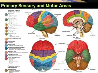
Somatosenses • Somatosensory pathway • Cell bodies in dorsal root ganglia • Spinothalamic tract (lateral) • Pain and temperature • Cross cord at entry level and ascend contralaterally • Dorsal columns • Precise touch • Ascend ipsilaterally until medulla, then crosses • Synapse in medial lemniscus of thalamus • Somatotopic primary somatosensory cortex on postcentral gyrus (contralateral to sensors).
Spinal Reflexes • Afferent (sensory) nerve enters cord thru dorsal root. • Synapses either directly on efferent nerve, or an intermediary (interneuron) in spinal cord. • Intermediaries can cross to opposite side. • Efferent (motor) nerve leaves ventral root. • Status sent to brain, which can modulate response.
Spinal Reflexes • Ex: Knee-jerk reflex (patellar tendon reflex) • Spinal reflexes normally act to keep flexors and extensors in balance (no motion). • The doctor’s hammer stretches the quadriceps tendon (as if knee was flexed). • Stretch receptors synapse in spinal cord on quadriceps motor neurons. • Quadriceps contract and knee extends to counter the “flexing” knee.
Neural Control of Movement • Several parts necessary for movement: • Sensory nerves (input) • Proprioceptors • Touch sensors • Vestibular sensors • Something to control the process: • Spinal (reflex) control of movement (involuntary) • Brain control of movement (voluntary) • Muscles (output)
Involuntary Motor Control • Reflexes • In spinal cord • Uni- or bi-lateral • Can be sensed and moderated by the cortex • Balance • Vestibular sensors • Vestibular nuceli (VN) of Medulla • Ventromedial pathways
Voluntary Motor Control • Motivation (what?) • Frontal lobe • Strategy (how?) • SMA, PMA, basal ganglia & posterior parietal cortices • Tactics (details) • Pre-motor cortex and cerebellum • Execution • Primary motor cortex, brainstem, spinal cord and muscles
Voluntary Motor Control • Voluntary movements • Thought originates in frontal cortex. • Posterior parietal area knows body position. • Descends to the basal ganglia (caudate nucleus, putamen and globus pallidus) for focusing. • Proceeds to premotor cortex for preplanning. • Cerebellum coordinates multiple muscles and is responsible for “motor memory.” • Then onto primary motor cortex on the precentral gyrus for final movement commands. • Lateral descending pathways to effector muscles.
Voluntary Motor Control Dopamine inhibits the basal ganglia from inhibiting movement.
The Cerebellum • Contains over 50% of CNS neurons! It must be important! • Cerebellum coordinates complex sequences of actions by many muscles. • Takes an idea of a motion apart, and returns details of force, direction and timing. • Lesions result in ataxias. Alcohol-induced cerebellar inhibition causes same uncoordinated and inaccurate movements.
The Cerebellum • Important site of motor learning • Consider ballistic motions - they are too fast for sensory information to be incorporated, so they must be based on estimations. • These estimations can only be made on the basis of experience, which must be adjusted and fine-tuned over time. • “Practice makes perfect”
Descending Pathways • Lateral pathway • Corticospinal and rubrospinal tracts • Voluntary movements controlled by the cortex • Ventromedial pathway • Tectospinal, vestibulospinal, reticulospinal tracts • Involuntary movements controlled by the brainstem • Balance & visual orientation