Download
1 / 21
220 likes | 401 Vues
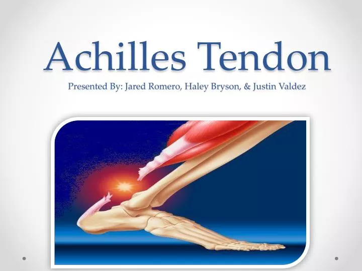
Achilles Tendon Presented By: Jared Romero, Haley Bryson, & Justin Valdez. Overview of Injury. Thickest tendon in the body. 1,2 Connects gastrocnemius, soleus, and plantaris to the calcaneus. 1 Commonly injured by sudden plantarflexion or dorsiflexion of ankle. 2. Risk Factors.

E N D
Achilles TendonPresented By: Jared Romero, Haley Bryson, & Justin Valdez
Overview of Injury • Thickest tendon in the body.1,2 • Connects gastrocnemius, soleus, and plantaris to the calcaneus.1 • Commonly injured by sudden plantarflexionor dorsiflexion of ankle.2
Risk Factors • Athletes, runners, basketball players. Older adults in high demand sports.1-15 • ~40 Years of Age1,3-5, 7, 15 • FluoroquinoloneAntiobiotics and Direct Steroid Injections into the tendon.3 • Previous Achilles Tendon injury.
Surgical Procedure • 2 Types of surgery to repair a torn Achilles.4-6 1) Open Surgery: Large, single incision.7 2) Percutaneous Surgery: surgeon makes several small incisions rather than one large one.8,9
Rehabilitation: Phase 1 • Surgery to 4 weeks after surgery. • Precautions: • Avoid long periods of dependent positioning of the foot.11 • Avoid excessive walking and standing • Rehab appointments are 1-2x per week • At end of phase 1: Goal is to have pain-free active dorsiflexion to 0 degrees.
Phase 1: Weeks 1-4 • ROM Exercises • Foot stabilized • Active ROM exercises for toes, knee, and hip. • Elevate Leg • Stretching • Stretching of the hamstrings, gastrocnemius, soleus and quad. • Straight Leg Lifts • Hip Abduction • Strengthening Isometric quad contractions Isometric knee extensions w/ ball
Phase 1: Cardiovascular Endurance • Stationary Cycle • With brace on, ride a stationary bike for 15-20 minutes a session if patient is able to without any pain. • Upper Body Ergometer • Continue to work on conditioning through the rehab process. 15-20 min a session.
Phase II Rehab: 4-8 weeks • Goals • Begin weight bearing11, encouraging normalized gait through all steps. 1-3, 11, 12, 16-18 • Increase ROM between 5° of dorsiflexion to 40° of plantar flexion1-3, 11, 12, 16-18 • Maintain Upper and Lower body strength, using exercises from phase I.1-3, 11, 12, 16-18 • Precautions • Slowly wean from the use of the boot11 • Avoid over stretching the repair1-3, 11, 12, 16-18 • Activities that have a high impact should be avoided • PROM should be gentle, do not push too far 1-3, 11, 12, 16-18 Rehab appointments are daily with an ATC; outpatient clinic is 1-2x/week.16-18
Phase II Rehab: 4-8 weeks • Treatment • If needed use of Grade I and II joint mobilizations at the talocrural joint, both anterior and posterior.16-18 • Use applicable modalities16-18 • Maintaining previous training levels • Stretching/ ROM • PROM at the foot and ankle16-18 • AROM dorsiflexion to 0°1-3, 11, 12, 16-18 • Once patient is partial weight bearing they may begin using a BAPS board while sitting to increase AROM.11
Phase II Rehab: 4-8 Weeks • Strengthening • Continue to strengthen all other areas of the body16-18 • 4-6 weeks isometric ankle exercises in all directions16-18 • 6-8 weeks add light resistance bands at the ankle in all directions 1-3, 11, 12, 16-18 • Cardiovascular Endurance/ Gait • As the patient becomes partial weight bearing begin walking in the pool.12 • Goal: normalized gait1-3, 11, 12, 16-18 • Once gait is normalized, power walking can be added for 30 mins per session. • Continue to use UBE and add the stationary bike • Work on proper gait technique, out of the water.12
Phase II Rehab: 4-8 weeks • Proprioception • Begin single leg stance on uninjured leg • Double leg standing with eyes closed when FWB permitted16-18 • Tandem stance on floor
Phase III: 8-12 Weeks • Goals • AROM between 15° of dorsiflexion and full plantar flexion16-18 • Single leg heel raise with good control for 10 seconds16-18 • Decrease pain with functional movements16-18 • Full weight bearing16-18 • Increase strength and endurance16-18 • Precautions • Limit forceful impact activities until approx. 12 weeks.1-3, 11, 12, 16-18 • Avoid activities where over compensation may occur1-3, 11, 12, 16-18 Rehab appointments are daily with ATC; 1x/ week outpatient setting16-18
Phase III: 8-12 Weeks • Stretching/ ROM • Use foam pad to increase ROM and stretch (proprioception) • Use grade III and IV posterior joint mobilizations as needed to increase mobility for dorsiflexion.1-3, 11, 12, 16-18 • Dorsiflexion door stretch (strength)16-18 • Strengthening • Continue the use of tubing in all directions, increase reps at first then increase resistance16-18 • Begin doing bilateral heel raises; start unilateral around 10 weeks1-3, 11, 12, 16-18 • Standing squats w/ ball • Lunges & Reverse Lunges
Phase III: 8-12 weeks • Cardiovascular Training: • Continue to use the stationary bike/ power walking in the pool (week 8) • 30 minute warm up per session • Progress to: • Elliptical (week 9) • Rowing (week 10) • Power walking on the treadmill or jogging in the pool (week 11) • Jogging may start at week 12.16-18
Phase III: 8-12 weeks • Proprioception • Tandem Stance • Balance board • Bosu ball tandem stance • Progress to single leg once single leg raise can be done:16-18 • Single leg stance • Single leg stance on bosu ball • Single leg stance on half foam roller
Phase 4: 12 weeks to completion of Rehab • Goals: • Jogging without pain for 2 miles. • Maintain regained ROM • Functional and sport specific movements with no pain. • Plyometrics without pain. • Return to play with no pain. • Precautions: • Ease into more impacted movements. • Progress only when pain is absent. • Avoid overcompensation.
Phase IV • Stretching/ROM • Full ROM should be present prior to this phase • Maintain ROM with slant board work in Phase IIIeach day prior to phase IV work. • Strengthening • Patient will progress from heel raises by adding ankle weights to increase resistance. • Continue with lunges but add weight as necessary
Phase IV • Plyometrics • Once able to complete weighted heel raises progress to double leg hops. • Progress to single leg hops. • Progress to lateral double leg hops. • Progress to lateral single leg hops. • Cardiovascular Endurance • Patient must be able to jog on a flat surface for up to 2 miles with no pain in order to progress to next level of running drills. • Weeks 12-14: strict jogging. • Weeks 14- finish: Sprinting will be added.
Phase IV • Speed • Body weight sprints of short yardage is required • build up to maximal effort by rehabs end • Sprints should be between 10 and 30 yards • Power • Power will be regained by taking simple movements such as the squat or a single leg skip and making it explosive • Squat jumps • Power skips • Agility • Figure 8 cone drills
References • Majewski M, Schaeren S, Kohlhaas U, Ochsner P. Postoperative rehabilitation after percutaneous Achilles tendon repair: Early functional therapy versus cast immobilization. Disability & Rehabilitation. October 30, 2008;30(20-22):1726-1732. • Suchak, Amar A., et al. Postoperative rehabilitation protocols for Achilles tendon ruptures: a meta-analysis. Clinical orthopaedics and related research 2006; 445: 216-221. • Amin N, Old A, Tabb L, Garg R, Toossi N, Cerynik D. Performance Outcomes After Repair of Complete Achilles Tendon Ruptures in National Basketball Association Players. American Journal Of Sports Medicine. August 2013;41(8):1864-1868. • Henríquez H, Muñoz R, Carcuro G, Bastías C. Is percutaneous repair better than open repair in acute achilles tendon rupture?. Clinical Orthopaedics & Related Research. April 2012;470(4):998-1003. • Hrnack S, Crates J, Barber F. Primary Achilles Tendon Repair with Mini-Dorsolateral Incision Technique and Accelerated Rehabilitation. Foot & Ankle International. October 2012;33(10):848-851. • Gigante A, Moschini A, Verdenelli A, Torto M, Ulisse S, Palma L. Open versus percutaneous repair in the treatment of acute Achilles tendon rupture: a randomized prospective study. Knee Surgery, Sports Traumatology, Arthroscopy. February 2008;16(2):204-209. • Willits K, Amendola A, Kirkley A, et al. Operative versus Nonoperative Treatment of Acute Achilles Tendon Ruptures: A Multicenter Randomized Trial Using Accelerated Functional Rehabilitation. Journal Of Bone & Joint Surgery, American Volume. December 2010;92-A(17):2767-2775. • Talbot J, Williams G, Bismil Q, Shaw D, Schilders E. Results of Accelerated Postoperative Rehabilitation Using Novel “Suture Frame” Repair of Achilles Tendon Rupture. Journal Of Foot & Ankle Surgery. March 2012;51(2):147-151.
References…Continued • LuiT. Endoscopic Achilles Tenolysis for Management of Heel Cord Pain after Repair of Acute Rupture of Achilles Tendon. Journal Of Foot & Ankle Surgery. 2013;52(1):125-127. • MaquirriainJ. Achilles tendon rupture: avoiding tendon lengthening during surgical repair and rehabilitation. The Yale Journal Of Biology And Medicine. September 2011;84(3):289-300. • SuchakA, Bostick G, Beaupré L, Durand D, Jomha N. The influence of early weight-bearing compared with non-weight-bearing after surgical repair of the Achilles tendon. Journal Of Bone & Joint Surgery, American Volume. September 2008;90(9):1876-1883. • Speck M, Klaue K. Early full weightbearing and functional treatment after surgical repair of acute Achilles tendon rupture. American Journal of Sports Medicine. 1998; 26(6): 789-793. • Lee S, Sileo M, McHugh M, et al. Cyclic Loading of 3 Achilles Tendon Repairs Simulating Early Postoperative Forces. American Journal Of Sports Medicine. April 2009;37(4):786-790. • Loudon J. Early Motion of the Ankle After Operative Treatment of a Rupture of the Achilles Tendon: A.. Physical Therapy. November 1999;79(11):1096. • GallasJ. Proprioceptive and strength deficits of the lower leg following Achilles tendon rupture and repair. Orthopaedic Physical Therapy Practice. December 2011;23(4):204-209. • Houlgum PA, Perrin DH, eds. Therapeutic Exercise For Musculoskeletal Injuries. Champaign, Il: Human Kinetics; 2010. • Massachusetts General Hospital. Achilles Tendon Repair Surgery Rehabilitation Protocol.http://www.massgeneral.org/ortho/services/sports/rehab/Achilles%20repair%20rehabilitation%20protocol.pdf. Accessed September 18, 2013 • University of Wisconsin Sports Medicine. Rehabilitation Guidelines for Achilles Tendon Repair. http://www.uwhealth.org/files/uwhealth/docs/sportsmed/SM-27399_AchillesTendonProtocol.pdf. Accessed September 18, 2013.


![ANCIENT GREECE [Map 05-01]](https://cdn3.slideserve.com/5762335/slide1-dt.jpg)