Download
1 / 56
560 likes | 565 Vues
Surveying the Chapter: Overview What We Have in Mind. Building blocks of the mind: neurons and how they communicate (neurotransmitters) Systems that build the mind: functions of the parts of the nervous system Supporting player: the slower-communicating endocrine system (hormones)

E N D
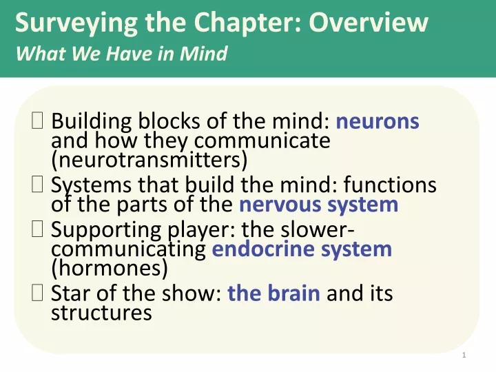
Surveying the Chapter: OverviewWhat We Have in Mind • Building blocks of the mind: neurons and how they communicate (neurotransmitters) • Systems that build the mind: functions of the parts of the nervous system • Supporting player: the slower-communicating endocrine system (hormones) • Star of the show: the brain and its structures
Searching for the self by studying the bodyPhrenology Phrenology (developed by Franz Gall in the early 1800’s): the study of bumps on the skull and their relationship to mental abilities and character traits • Phrenology yielded one big idea--that the brain might have different areas that do different things (localization of function).
Today’s search for the biology of the self: biological psychology • Biological psychology includes neuroscience, behavior genetics, neuropsychology, and evolutionary psychology. • All of these subspecialties explore different aspects of: how the nature of mind and behavior is rooted in our biological heritage. • Our study of the biology of the mind begins with the “atoms” of the mind: neurons.
Neurons and Neuronal Communication:The Structure of a Neuron There are billions of neurons (nerve cells) throughout the body.
Crash Course Psych • Crash Course-Neurons
Action potential:a neural impulse that travels down an axon like a wave Just as “the wave” can flow to the right in a stadium even though the people only move up and down, a wave moves down an axon although it is only made up of ion exchanges moving in and out.
When does the cell send the action potential?... when it reaches a threshold How neurons communicate(with each other): The neuron receives signals from other neurons; some are telling it to fire and some are telling it not to fire. • When the threshold is reached, the action potential starts moving. • Like a gun, it either fires or it doesn’t; more stimulation does nothing. • This is known as the “all-or-none” response. The action potential travels down the axon from the cell body to the terminal branches. The signal is transmitted to another cell. However, the message must find a way to cross a gap between cells. This gap is also called the synapse. The threshold is reached when excitatory (“Fire!”) signals outweigh the inhibitory (“Don’t fire!”) signals by a certain amount.
The Synapse The synapse is a junction between the axon tip of the sending neuron and the dendrite or cell body of the receiving neuron. The synapse is also known as the “synaptic junction” or “synaptic gap.”
Neurotransmitters Neurotransmitters are chemicals used to send a signal across the synaptic gap.
Reuptake:Recycling Neurotransmitters [NTs] Reuptake: After the neurotransmitters stimulate the receptors on the receiving neuron, the chemicals are taken back up into the sending neuron to be used again.
Neural Communication: Seeing all the Steps Together
Dopamine pathways Serotonin pathways Networks of neurons that communicate with dopamine are involved in focusing attention and controlling movement. Networks of neurons that communicate with serotonin help regulate mood.
Hearing the messageHow Neurotransmitters Activate Receptors When the key fits, the site is opened.
Keys that almost fit:Agonist and AntagonistMolecules An antagonist molecule fills the lock so that the neurotransmitter cannot get in and activate the receptor site. An agonistmolecule fills the receptor site and activates it, acting like the neurotransmitter.
The Inner and Outer Parts of the Nervous System The central nervous system [CNS] consists of the brain and spinal cord. The CNS makes decisions for the body. The peripheral nervous system [PNS] consists of ‘the rest’ of the nervous system. The PNS gathers and sends information to and from the rest of the body.
The AutonomicNervous System: The sympathetic NS arouses(fight-or-flight)The parasympatheticNS calms(rest and digest)
The Body’s “Slow but Sure” Endocrine Message System • The endocrine system sends molecules as messages, just like the nervous system, but it sends them through the bloodstream instead of across synapses. • These molecules, called hormones, are produced in various glands around the body. • The messages go to the brain and other tissues. The endocrine system refers to a set of glands that produce chemical messengers called hormones.
produce hormones such as adrenaline/epinephrine, noradrenaline/norepinephrine, and cortisol. Adrenal Glands Adrenal Glands • The sympathetic “fight or flight” nervous system responds to stress by sending a message to adrenal glands to release the hormones listed above. • Effect: increased heart rate, blood pressure, and blood sugar. These provide ENERGY for the fight or flight! Pancreas
The Pituitary Gland • The pituitary gland is the “master gland” of the endocrine system. • It is controlled through the nervous system by the nearby brain area--the hypothalamus. • The pituitary gland produces hormones that regulate other glands such as the thyroid. • It also produces growth hormone (especially during sleep) and oxytocin, the “bonding” hormone. Pituitary gland
Investigating the Brain and Mind: Strategies for finding out what is different about the mind when part of the brain isn’t working normally: • case studies of accidents (e.g. Phineas Gage) • case studies of split-brain patients (corpus callosum cut to stop seizures) • lesioning brain parts in animals to find out what happens • chemically numbing, magnetically deactivating, or electrically stimulating parts of the brain How did we move beyond phrenology and get inside the skull and under the “bumps”? • by finding what happens when part of the brain is damaged or otherwise unable to work properly • by looking at the structure and activity of the brain: CAT, MRI, fMRI, and PET scans
Studying cases of brain damage When a stroke or injury damages part of the brain, we have a chance to see the impact on the mind.
Intentional brain damage: Lesions (surgical destruction of brain tissue) • performed on animals • has yielded some insights, especially about less complex brain structures • no longer necessary, as we now can chemically or magnetically deactivate brain areas to get similar information
Split-Brain Patients • “Split” = surgery in which the connection between the brain hemispheres is cut in order to end severe full-brain seizures • Study of split-brain patients has yielded insights discussed at the end of the chapter
We can stimulate parts of the brain to see what happens • Parts of the brain, and even neurons, can be stimulated electrically, chemically, or magnetically. • This can result in behaviors such as giggling, head turning, or simulated vivid recall. • Researchers can see which neurons or neural networks fire in conjunction with certain mental experiences, and even specific concepts.
EEG: electroencephalogram PET: positron emission tomography An EEG (electroencephalogram) is a recording of the electrical waves sweeping across the brain’s surface. It is useful in studying seizures and sleep. The PET scan allows us to see what part of the brain is active by tracing where a radioactive form of glucose goes while the brain performs a given task.
MRI: magnetic resonance imaging fMRI: functional MRI Functional MRI reveals brain activity and function rather than structures. MRI (magnetic resonance imaging) makes images from signals produced by brain tissue after magnets align the spin of atoms. The arrows below show ventricular enlargement in a schizophrenic patient (right). Functional MRI compares successive MRI images taken a split second apart, and shows changes in the level of oxygen in bloodflow in the brain.
The Brainstem: Pons and Medulla • The medulla controls the most basic functions such as heartbeat and breathing. • Someone with total brain damage above the medulla could still breathe independently, but someone with damage in this area could not. • The pons helps coordinate automatic and unconscious movements.
The Thalamus (“Inner Chamber”) • The thalamus is the “sensory switchboard” or “router.” • All sensory messages, except smell, are routed through the thalamus on the way to the cortex (higher, outer brain). • The thalamus also sends messages from the cortex to the medulla and cerebellum.
Reticular (“Netlike”) Formation • The reticular formation is a nerve network in the brainstem. • It enables alertness, (arousal) from coma to wide awake (as demonstrated in the cat experiments). • It also filters incoming sensory information.
Cerebellum (“little brain”) The cerebellum helps coordinate voluntary movement such as playing a sport. The cerebellum has many other functions, including enabling nonverbal learning and memory.
The Limbic (“Border”) System The limbic system coordinates: • emotions such as fear and aggression. • basic drives such as hunger and sex. • the formation of episodic memories. The hippocampus (“seahorse”) • processes conscious, episodic memories. • works with the amygdala to form emotionally charged memories. The Amygdala (“almond”) • consists of two lima bean- sized neural clusters. • helps process emotions, especially fear and aggression.
The Amygdala • Electrical stimulation of a cat’s amygdala provokes aggressive reactions. • If you move the electrode very slightly and cage the cat with a mouse, the cat will cower in terror.
The Hypothalamus: Thalamus • lies below (“hypo”) the thalamus. • regulates body temperature and ensures adequate food and water intake (homeostasis), and is involved in sex drive. • directs the endocrine system via messages to the pituitary gland. The Hypothalamus as a Reward Center Riddle: Why did the rat cross the grid? Why did the rat want to get to the other side? Pushing the pedal that stimulated the electrode placed in the hypothalamus was much more rewarding than food pellets.
The Cerebral Cortex The lobes consist of: • outer grey “bark” structure that is wrinkled in order to create more surface area for 20+ billion neurons. • inner white stuff—axonslinking parts of the brain. • 180+ billion glial cells, which feed and protect neurons and assist neural transmission. 300 billion synaptic connections The brain has left and right hemispheres
The Lobes of the Cerebral Cortex: Preview involved in speaking and muscle movements and in making plans and judgments • Frontal Lobes • Parietal Lobes • Occipital Lobes • Temporal Lobes include the sensory cortex include the visual areas; they receive visual information from the opposite visual field include the auditory processing areas
Functions of the Brain: The Motor and Sensory Strips Output: Motor cortex (Left hemisphere section controls the body’s right side) Input: Sensory cortex (Left hemisphere section receives input from the body’s right side) Axons receiving motor signals FROM the cortex Axons sending sensory information TO the cortex
Sensory Functions of the Cortex • Thesensory stripdeals with information from touch stimuli. • The occipital lobe deals with visualinformation. • Auditoryinformation is sent to thetemporal lobe.
The Visual Cortex This fMRI scan shows increased activity in the visual cortex when a person looks at a photograph.
Association function of the cortex More complex animals have more cortical space devoted to integrating/associating information
Association Areas:Frontal Lobes • The frontal lobes are active in “executive functions” such as judgment, planning, and inhibition of impulses. • The frontal lobes are also active in the use of working memory and the processing of new memories.
Phineas Gage (1823-1860) Case study: In a work accident, a metal rod shot up through Phineas Gage’s skull, destroying his eye and part of his frontal lobes. After healing, he was able to function in many ways, but his personality changed; he was rude, odd, irritable, and unpredictable. Possible explanation: Damage to the frontal lobes could result in loss of the ability to suppress impulses and to modulate emotions.
Parietal Lobe Association Areas This part of the brain has many functions in the association areas behind the sensory strip: • managing input from multiple senses • performing spatial and mathematical reasoning • monitoring the sensation of movement
Temporal Lobe Association Areas Some abilities managed by association areas in this “by the temples” lobe: • recognizing specific faces • managing sensory input related to sound, which helps the understanding of spoken words
Whole-brain Association Activity Whole-brain association activity involves complex activities which require communication among association areas across the brain such as: • memory • language • attention • meditation and spirituality • consciousness
Specialization and Integration Five steps in reading a word aloud:
Plasticity: The Brain is Flexible If the brain is damaged, especially in the general association areas of the cortex: • the brain does not repair damaged neurons, BUT it can restore some functions • it can form new connections, reassign existing networks, and insert new neurons, some grown from stem cells This 6-year-old had a hemispherectomy to end life-threatening seizures; her remaining hemisphere compensated for the damage.