Download
1 / 74
830 likes | 1.18k Vues
Amino Acids, Peptides, and Proteins. Proteins Occur in every living organism Are of many different types Have many different biological functions Keratin of skin and fingernails Fibroin of silk and spider webs

E N D
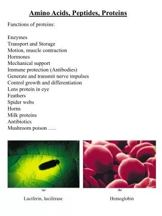
Amino Acids, Peptides, and Proteins Proteins • Occur in every living organism • Are of many different types • Have many different biological functions • Keratin of skin and fingernails • Fibroin of silk and spider webs • Estimated 50,000 to 70,000 enzymes that catalyze the biological functions of the human body • Made up of many amino acids linked together
Amino Acids, Peptides, and Proteins Amino acids are difunctional • Contain both a basic amino group and an acidic carboxyl group • Can join together into long chains by forming bonds between the –NH2 of one amino acid and the –CO2H of another • Peptides are chains with fewer than 50 amino acids • Proteins are large chains of amino acids
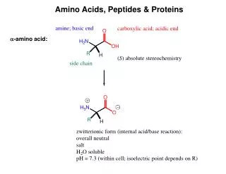
19.1 Structures of Amino Acids Amino acids exist in aqueous solution primarily in the form of a dipolar ion, or zwitterion Zwitterion • German zwitter, meaning “hybrid” • A neutral dipolar molecule in which the positive and negative charges are not adjacent
Amino acid zwitterions Are internal salts Have large dipole moments Are soluble in water but are insoluble in hydrocarbons Are crystalline substances with relatively high melting points Are amphiprotic Can react as acids Reaction takes place in an aqueous base solution Loses a proton to form an anion Can react as bases Reaction takes place in an aqueous acid solution Accepts a proton to yield a cation Structures of Amino Acids
Structures of Amino Acids All 20 amino acids found in proteins are a-amino acids • The amino group in each is a substituent on the a carbon atom – the one next to the carbonyl group • 19 of the amino acids are primary amines, RNH2 • Differ in the side chain, the substituent attached to the a carbon • Proline, a secondary amine, is the only amino acid whose nitrogen and a carbon atoms are part of the ring
Structures of Amino Acids Selenocysteine and pyrrolysine are amino acids found in some organisms
Structures of Amino Acids More than 700 nonprotein amino acids are found in nature • g-Aminobutyric acid (GABA) is found in the brain and acts as a neurotransmitter • Homocysteine is found in blood and is linked to coronary heart disease • Thyroxine is found in the thyroid gland, where it acts as a hormone
Structures of Amino Acids a Carbons of amino acids are chirality centers, except for glycine, H2NCH2CO2H • Nature uses only one enantiomer to build proteins • Often referred to as L amino acids • The nonnaturally occurring enantiomers are called the D amino acids
Structures of Amino Acids The 20 common amino acids can be acidic, basic, or neutral, depending on their side chains • 2 (aspartic acid and glutamic acid) of the 20 have an extra carboxylic acid function in their side chains • 3 (lysine, arginine, and histidine) have basic amino groups in their side chains • 15 have neutral side chains • Cysteine, a thiol, and tyrosine, a phenol, have weakly acidic side chains that can be deprotonated in a sufficiently basic solution
Structures of Amino Acids At physiological pH 7.3 within cells: • The side-chain carboxyl groups of aspartic acid and glutamic acid are deprotonated • The basic side-chain nitrogens of lysine and arginine are protonated • Histidine contains a heterocyclic imidazole ring in its side chain and is not basic enough to be protonated • Only the pyridine-like nitrogen is basic • The pyrrole-like nitrogen is nonbasic because its lone pair of electrons is part of the 6p electron aromatic imidazole ring
19.2 Amino Acids and the Henderson- Hasselbalch Equation: Isoelectric Points Using the Henderson-Hasselbalch equation the ratio of [A-] to [HA] in the solution can be calculated
Amino Acids and the Henderson-Hasselbalch Equation: Isoelectric Points Species present in a 1.00 M solution of alanine at pH = 9.00 • From Table 19.1 • [+H3NCH(CH3)CO2H] has pKa(a-CO2H) = 2.34 • [+H3NCH(CH3)CO2-] has pKa(a-NH3+) = 9.69
Amino Acids and the Henderson-Hasselbalch Equation: Isoelectric Points so also
Amino Acids and the Henderson-Hasselbalch Equation: Isoelectric Points Solving the two simultaneous equations gives [HA] = 0.83 and [A-] = 0.17 Therefore, in a 1.00 M solution at pH = 9.00 83% of alanine molecules are neutral zwitterionic [+H3NCH(CH3)CO2-] 17% of alanine molecules are deprotonated [+H3NCH(CH3)CO2H]
Amino Acids and the Henderson-Hasselbalch Equation: Isoelectric Points Similar calculations at other pH values leads to titration curve
Amino Acids and the Henderson-Hasselbalch Equation: Isoelectric Points In acid solution, an amino acid is protonated and exists primarily as a cation In basic solution, an amino acid is deprotonated and exists primarily as an anion • Isoelectric point, pI • The pH at which the number of positive charges and the number of negative charges on a protein or an amino acid are equal • Depends on the structure of the amino acid • Values given in Table 19.1
Amino Acids and the Henderson-Hasselbalch Equation: Isoelectric Points The pIof any amino acid is the average of the two acid-dissociation constants that involve the neutral zwitterion • The 4 amino acids with either a strongly or weakly acidic chain, pIis the average of the two lowest pKa values • The 13 amino acids with a neutral side chain, pI is the average of pKa1 and pKa2 • The 3 amino acids with a basic side chain, pI is the average of the two highest pKa values
Amino Acids and the Henderson-Hasselbalch Equation: Isoelectric Points Proteins have an overall pI because of the acidic or basic amino acids they may contain • Lysozyme has a preponderance of basic amino acids and thus has a high isoelectric point (pI = 11.0) • Pepsin has a preponderance of acidic amino acids and a low isoelectric point (pI ~ 1.0) The solubilities and properties of proteins with different pI ’s are strongly affected by the pH of the medium • Solubility in water is usually lowest at the isoelectric point where the protein has no net charge, and higher above and below the pI, where the protein is charged
Amino Acids and the Henderson-Hasselbalch Equation: Isoelectric Points Electrophoresis uses isoelectric points to separate a mixture of proteins into its pure constituents • A mixture of proteins is placed near the center of a strip of paper or gel • The paper or gel is moistened with an aqueous buffer of a given pH • Electrodes are connected to the ends of the strip and an electric potential is applied • Proteins separate according to charges (determined by the pH of the buffer strip) • Proteins with negative charges migrate slowly toward the positive electrode • Proteins with positive charges migrate slowly toward the negative electrode Different proteins migrate at different rates depending upon their pI and the pH of the buffer
19.3 Synthesis of Amino Acids The Amidomalonate Synthesis • Used for synthesizing a-amino acids: Amidomalonate synthesis of amino acids is an extension of the malonic ester synthesis • Conversion of diethyl acetamidomalonate into an enolate ion by treatment with a base • SN2 alkylation with a primary alkyl halide • Hydrolysis of both the amide group and the esters occurs when the alkylated product is warmed with aqueous acid • Decarboxylation takes place to yield an a-amino acid Preparation of aspartic acid from ethyl bromoacetate, BrCH2CO2Et • Reaction yields a racemate
Synthesis of Amino Acids Reductive Amination of a-Keto Acids • Another method of synthesizing a-amino acids • Reduces an a-keto acid with ammonia and a reducing agent • Preparation of alanine by treatment of pyruvic acid with ammonia in the presence of NaBH4 • Reaction proceeds through formation of an intermediate imine that is then reduced • Reaction yields a racemate
Synthesis of Amino Acids Enantioselective Synthesis • One of the two methods used to obtain enantiomerically pure amino acids • More efficient than resolving the racemate into its pure enantiomers • Prepares only the desired S enantiomer • Idea of the synthesis is to find a chiral reaction catalyst that will temporarily hold a substrate molecule in an unsymmetrical environment • While in the unsymmetrical environment, the substrate may be more open to reaction on one side than on another, leading to an excess of one enantiomeric product over another
Synthesis of Amino Acids William Knowles discovered that a-amino acids can be prepared enantioselectively by hydrogenation of a Z enamido acid with a chiral rhodium hydrogenation catalyst • (S)-Phenylalanine, is prepared in 98.7% purity contaminated by only 1.3% if the (R) enantiomer when a chiral rhodium catalyst is used • Knowles received the 2001 Nobel Prize in Chemistry
Synthesis of Amino Acids Most effective catalysts for enantioselective amino acid synthesis are coordination complexes of rhodium(I) with cyclooctadiene (COD) and a chiral diphosphine such as (R,R)-1,2-bis(o-anisylphenylphosphine)ethane, the DiPAMP ligand
19.4 Peptides and Proteins Peptides and proteins are amino acid polymers Residues • An amino in a protein chain • Joined together by amide bonds, or peptide bonds An amino group from one residue forms an amide bond with the carboxyl group of a second residue • Alanylserine is the dipeptide that results when an amide bond is formed between the alanine carboxyl and the serine amino group
Peptides and Proteins Two dipeptides can result from reaction between alanine and serine, depending on which carboxyl group reacts with which amino group • If the alanine amino group reacts with the serine carboxyl, serylalanine results
Peptides and Proteins Proteins • The protein’s backbone is continuous chain of atoms running the length of a protein or other polymer • The repetitive sequence of –N–CH–CO–atoms Polypeptides • Convention for writing peptides: • N-terminal amino acid on the left • Peptides with the free –NH2 group • C-terminal amino acid on the right • Peptides with the free –CO2H group • The name of the peptide is indicated using abbreviations • Alanylserine is abbreviated Ala-Ser or A-S • Serylalanine is abbreviated Ser-Ala or S-A
Peptides and Proteins Amide Bonds • Amide nitrogens are nonbasic because their unshared electron pair is delocalized by interaction with the carbonyl group • The overlap of the nitrogen p orbital with the p orbital of the carbonyl group imparts a certain amount of double-bond character to the C-N bond and restricts rotation around it • The amide bond is planar • The N-H is oriented 180º to the C=O
Peptides and Proteins A disulfide linkage, RS-SR • A covalent bond in peptides • Formed between two cysteine residues • Links chains of peptides together • Creates loops within a single chain of peptides • Vasopressin, an antidiuretic hormone found in the pituitary gland Note: the C-terminal end of vasopressin occurs as the primary amide, -CONH2
19.5 Amino Acid Analysis of Peptides Determination of structure of a protein or peptide • What amino acids are present? • How much of each is present? • In what sequence do the amino acids occur? Amino acid analyzer is an automated instrument that provides the answers to the first two questions • The peptide is broken into its constituent amino acids • Reducing all disulfide bonds • Capping the –SH groups of cysteine residues by SN2 reaction with iodoacetic acid • Hydrolyzing the amide bonds by heating with aqueous 6 M HCl at 110 ºC for 24 hours • The resultant amino acid mixture is analyzed by: • High-pressure liquid chromatography (HPLC) • Ion-exchange chromatography
Amino Acid Analysis of Peptides • As each amino acid exits (elutes) from the end of the ion-exchange chromatography column, it reacts with a solution of ninhydrin, giving an intense purple color • The color is detected by a spectrometer and a plot of elution time versus spectrometer absorbance is obtained
The identities of the amino acids in a peptide can be determined simply by noting the various elution times The amount of each amino acid in the sample is determined by measuring the intensity of the purple color resulting from its reaction with ninhydrin The results of amino acid analysis of a standard equimolar mixture of 17a-amino acids Amino acid analysis requires about 100 picomoles (2-3 mg) of sample for a protein containing about 200 residues Amino Acid Analysis of Peptides
19.6 Peptide Sequencing: The Edman Degradation With the known identities and amounts of amino acids, the peptide is sequenced to find out in what order the amino acids are linked together • Methods of peptide sequencing: • Mass spectrometry using electrospray ionization (ESI) • Mass spectrometry using matrix-assisted laser desorption ionization (MALDI) linked to a time-of-flight (TOF) mass analyzer • Edman degradation • Cleave one amino acid at a time from an end of the peptide chain • Cleaved amino acid is separated and identified • Repeated until entire peptide sequence is known
Peptide Sequencing: The Edman Degradation • Automated protein sequencers • Allow as many as 50 repetitive sequencing cycles to be carried out before a buildup of unwanted by-products interferes with the results • Provide information from as little as 1 to 5 picomoles of sample – less than 0.1 mg
Peptide Sequencing: The Edman Degradation • Edman degradation for N-terminal analysis peptides • Involves treatment of a peptide with phenyl isothiocyanate (PITC), C6H5–N=C=S, followed by treatment with trifluoroacetic acid • The phenylthiohydantoin (PTH) is identified chromatographically by comparison of its elution times with the known elution times of PTH derivatives of all 20 common amino acids
Peptide Sequencing: The Edman Degradation Partial hydrolysis of a peptide can carried out either chemically, using aqueous acid, or enzymatically • Acidic hydrolysis is unselective and leads to mixtures of small fragments • Enzymatic hydrolysis is quite specific • Trypsin catalyzes hydrolysis at the carboxyl side of the basic amino acids arginine and lysine • Chymotrypsin cleaves only at the carboxyl side of the aryl-substituted amino acids phenylalanine, tyrosine, and tryptophan
19.7 Peptide Synthesis During the course of a peptide synthesis, many different amide bonds must be formed in a specific order • The solution of the specificity problem is to protect some functional groups rendering them unreactive while leaving exposed only those functional groups wanted for reaction • To synthesize Ala-Leu, by coupling alanine with leucine • Protect the –NH2 group of alanine and the –CO2H group of leucine to render them unreactive • Form the desired amide bond • Remove the protecting groups
Peptide Synthesis Amino- and carboxyl-protecting groups • Carboxyl groups are often protected by converting them into methyl or benzyl esters • Both groups are easily introduced by standard methods of ester formation • Both groups are easily removed by mild hydrolysis with aqueous NaOH • Benzyl esters can also be cleaved by catalytic hydrogenolysis of the weak benzylic C-O bond (RCO2–CH2Ph + H2 PhCH3)
Peptide Synthesis • Amino groups are often protected as their tert-butoxycarbonyl amide, or Boc, derivatives • Protecting group is introduced by reaction of the amino acid with di-tert-butyl dicarbonate in a nucleophile acyl substitution reaction • Protecting group is removed by brief treatment with a strong organic acid such as trifluoroacetic acid, CF3CO2H
Peptide Synthesis Five steps are needed to synthesize a dipeptide such as Ala-Leu using the Boc protecting group
Merrifield solid-phase method simplifies the synthesis of a large peptide chain Peptide synthesis is carried out with the growing amino acid chain covalently bonded to small beads of polymer resin In the original Merrifield procedure, polystyrene resin was used 1 of every 100 or so benzene rings contained a chloromethyl (-CH2Cl) group A Boc-protected C-terminal amino acid was bonded to the resin through an ester bond formed by SN2 reaction Peptide Synthesis
Peptide Synthesis • With the first amino acid bonded to resin, a repeating series of four steps is carried out to build a peptide
Peptide Synthesis The most commonly used resins at present are the Wang resin or the PAM (phenylacetamidomethyl) resin • The most commonly used N-protecting group is the fluorenylmethyloxycarbonyl, or Fmoc group
Peptide Synthesis Robotic, computer-controlled peptide synthesis used to automatically repeat the coupling, washing, and deprotection steps with different amino acids • Each step occurs in high yield • The peptide intermediates are never removed from the insoluble polymer until the final step • Using this procedure, up to 30 mg of a peptide with 20 amino acids can be routinely prepared