Download
1 / 10
330 likes | 1.49k Vues
Equine Calcaneal Bursitis. Christina Copple, DVM Radiology Resident Monday September 21, 2009 Accession #’s 120984 & 120974. “Freck” 16yr old Arabian gelding. Intermittent left hind lame Effusion at lateral aspect of right hock (tarsus) and reluctant to pick up right foot

E N D
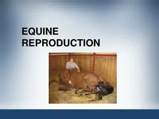
Equine Calcaneal Bursitis Christina Copple, DVM Radiology Resident Monday September 21, 2009 Accession #’s 120984 & 120974
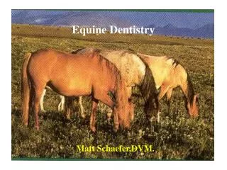
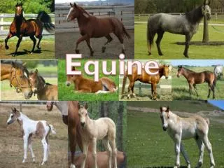
“Freck” 16yr old Arabian gelding • Intermittent left hind lame • Effusion at lateral aspect of right hock (tarsus) and reluctant to pick up right foot • Moderate to severe swelling over tuber calcaneus of left hock (tarsus) http://www.horse-wallpaper.com/backgrounds/white-arabian-horse.jpg http://media.photobucket.com/image/swollen%20equine%20hock%20joint/Glacierpt/Hanyshock002.jpg
Anatomy of Tarsus and Calcaneal Bursa 1 – Superficial Digital Flexor Tendon (SDFT) 1’ – Calcaneal Bursa 2 – Gastrocnemius 4 & 2’ – Tuber Calcaneus Dyce, et al. Textbook of Veterinary Anatomy 3rd ed. 2002. pg 614 & pg 615
Radiograph of Left Tarsus Swelling and increased soft tissue opacity in region of calcaneal bursa Lateromedial projection
Radiograph of Left Tarsus Two irregular & ill-defined lucencies in plantaromedial aspect of tuber calcaneus Dorsoplantar flexed projection
Ultrasound of left calcaneal bursa, tuber calcanei and associated soft tissue structures SDFT Anechoic fluid-filled calcaneal bursa with thickened synovial lining Gastrocnemius tendon
Ultrasound of left calcaneal bursa, tuber calcanei and associated soft tissue structures Defect in medial margin of SDFT with out pocketing of synovium into SQ tissues Tuber calcaneus
Ultrasound of left calcaneal bursa, tuber calcanei and associated soft tissue structures Defects in periosteum and irregular surface of tuber calcaneus
Septic Calcaneal Bursitis • Synovial fluid analysis – consistent with severe suppurative inflammation • Arthroscopy of left calcaneal bursa for lavage and debridement • Trauma results in hemorrhage and inflammation • Penetrating wounds are common • Sequela – tendon adhesions within bursa
References • Dyce, K.M., DVM&S, BSc, MRCVS, W.O. Sack, DVM, PhD, Dr.med.vet, and C.J.G. Wensing, DVM, PhD. Textbook of Veterinary Anatomy 3rd ed. Saunders. Philadelphia, PA. 2002. • Reef, Virginia B., DVM. Equine Diagnostic Ultrasound. Saunders. Philadelphia, PA. 1998.