Download
1 / 2
30 likes | 208 Vues
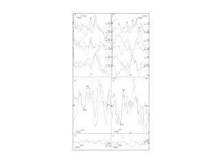
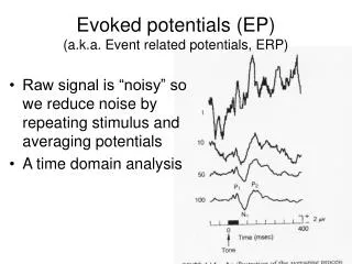
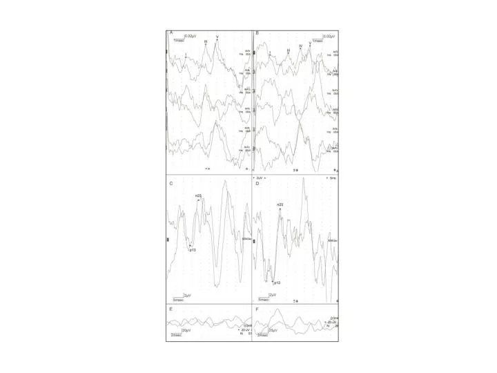
Supplemental Digital Content 2. Brainstem auditory evoked potentials with left ear (A) and right ear (B) stimulation in relation to our first case and the MRI shown in figure 1. The top trace in each figure represents the ipsilateral ear - Cz derivation,

E N D
Supplemental Digital Content 2. Brainstem auditory evoked potentials with left ear (A) and right ear (B) stimulation in relation to our first case and the MRI shown in figure 1. The top trace in each figure represents the ipsilateral ear -Cz derivation, the middle trace is ipsilateral - contralateral ear, and the bottom trace is contralateral ear – Cz. The left ear responses are within normal limits, but right ear stimulation resulted in abnormal waveform morphologies. Sweep was 1 msec/division, sensitivity was 0.05 uV/division. Cervical vestibular evoked potentials from our first case. (C) and (D) represent the responses from stimulating the left and right ear respectively. The bottom traces represent the parallel recording or rectified EMG from the same recording electrodes on the left (E) and on the right (F). The cVEMPs were within normal limits bilaterally. For the cVEMPs, the sweep was 5 msec/division, sensitivity was 5 uV/division. For the rectified EMG activity, the sweep was 2 msec/division and the sensitivity was 20 uV/division. A high background level of muscle activity is superimposed on the cVEMP response, more than usual when compared to figures 4 and 6, as this patient had difficulty in maintaining sternocleidomastoid muscle contraction.