Download
1 / 63
650 likes | 810 Vues
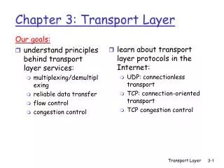
CHAPTER 1 - TRANSPORT. BIOLOGY FORM 5. In vertebrates, blood vessels from a tubular network Arteries carry blood away from the heart Veins return blood to the heart Capillaries connect arteries to veins

E N D
CHAPTER 1 - TRANSPORT BIOLOGY FORM 5
In vertebrates, blood vessels from a tubular network • Arteries carry blood away from the heart • Veins return blood to the heart • Capillaries connect arteries to veins • As blood plasma passes through capillaries, pressure forces fluid out of the capillary walls • Some of this interstitial fluid returns directly to capillaries • Some enters lymph vessels • This lymph is returned to venous blood at specific sites
Functions of Vertebrate Circulatory Systems • 1. Transportation • Respiratory • Transport O2 to cells for aerobic respiration • Transport CO2 to lungs/gills for elimination • Nutritive • Transport of absorbed products of digestion to cells • Excretory • Metabolic wastes and excessive water are filtered in the kidney and excreted in urine
2. Regulation • Hormones are transported from endocrine glands to distant target organs • Help maintain a constant body temperature in homeotherms • 3. Protection • Blood clotting protects against blood loss • White blood cells provide immunity against many-disease causing agents
Fig. 25.8 The flow of blood 25.2 Architecture of the VertebrateCirculatory System • The cardiovascularsystem of vertebrates consists of • 1. Heart • Pump • 2. Blood vessels • Network of tubes • 3. Blood • Circulating fluid
Fig. 25.3 The capillary network connects arteries with veins • Blood loses most of its pressure and velocity as it passes through the vast capillary network
Fig. 25.4a Arteries: Highways from the Heart • Blood comes from the heart in large pulses • Thus the artery must be able to expand • Arterial walls are made up of three layers • Arterioles are smaller in diameter than arteries • Their surrounding muscle layer can be relaxed to enlarge diameter
Fig. 25.4b Capillaries: Where Exchange Takes Place • Transport oxygen and nutrients from blood to body’s cells and pick up carbon dioxide • They have thin walls to allow diffusion to take place • Individual capillaries have high resistance to flow • But the total cross-sectional area of capillaries is greater than that of arteries leading to it
Fig. 25.4c Vein Artery Fig. 25.6 Veins: Returning Blood to the Heart • Walls have thinner layers of muscle and elastic fiber than arteries • When empty, walls collapse
Fig. 25.7 Veins: Returning Blood to the Heart • Blood flow back to the heart is aided by • 1. Low pressure in veins • 2. Skeletal muscles • 3. Unidirectional valves
Fig. 25.9 1.4 The Lymphatic System:Recovering Lost Fluid • The cardiovascular system is very leaky • To collect and recycle leaked fluid, the body uses a second circulatory system called the lymphatic system
Fig. 25.10 • Blood pressure forces fluid out of capillaries • Most of this interstitial fluid returns by osmosis • Excess fluid is drained into lymphatic capillaries • In the lymphatic system the fluid is called lymph
Lymphatic vessels contain a series of one-way valves • Permit movement only in the direction of the neck • The lymphatic system has three important functions • 1. Returns proteins to circulation • If proteins are not returned to the blood, a condition called edema (body swelling) results • 2. Transports fats absorbed from the intestine • Lymph capillaries, called lacteals, absorb fats from the small intestine • 3. Aids in the body’s defense • Lymph nodesare filled with white blood cells
25.4 Blood • Blood comprises about 5% of body mass • It is composed of • A fluid called plasma • Several different kinds of cells
Blood Plasma: The Body’s Fluid • Blood plasmais a complex solution of water and • 1. Metabolites and wastes • Glucose, vitamins, hormones and wastes • 2. Salts and ions • Chief plasma ions: sodium, chloride, bicarbonate • Minor ions: calcium, magnesium, copper • 3. Proteins • Act as an osmotic counterforce • Major protein: serum albumin • Other proteins: fibrinogen and antibodies
Blood Cells Circulate Through the Body • The fraction of blood volume that is occupied by cells is termed the blood’s hematocrit • In humans it is usually about 45% • The three principal types of blood cells are • Erythrocytes • Leukocytes • Platelets
Erythrocytes (red blood cells) • Carry hemoglobin, and therefore, oxygen to cells • Do not contain a nucleus • Leukocytes (white blood cells) • Defend the body against microbes and foreign substance • Neutrophils • Monocytes/Macrophages • Lymphocytes • B cells – Produce antibodies • T cells – Drill holes in invading bacteria
Fig. 25.11 • Platelets • Cell fragments that are bits of the cytoplasm of large bone marrow cells called megakaryocytes • Do not contain a nucleus • Play a key role in blood clotting • Stimulate the formation of fibrin from fibrinogen Fibrin
Fig. 25.13a 25.5 Fish Circulation • The fish heart is a tube consisting of four chambers • Sinus venosus and atrium, are collection chambers • Ventricle and conus arteriosus, are pumping chambers
Fig. 25.13b • The heart beat in fishes has a peristaltic sequence • Starts at the rear (SV) and moves to the front • Gill respiration provides fully oxygenated blood to the body • However, circulation is sluggish • This limits rate of oxygen delivery to rest of body
25.6 Amphibian and ReptileCirculation • The advent of lungs resulted in two circulations • 1. Pulmonary circulation • Delivers blood to the lungs • 2. Systemic circulation • Delivers blood to the rest of the body
Fig. 25.14a • The amphibian heart has two structural features that reduce mixing of oxygenated & deoxygenated blood • 1. The atrium is divided into two chambers by a septum • 2. Conus arteriosus is partially separated by another septum • Amphibians in water supplement the oxygenation of blood by a process called cutaneous respiration
Fig. 25.14b • Among reptiles, additional modifications have further reduced the mixing of blood in the heart • The ventricle is partially divided into two chambers by a septum • The separation is complete in the crocodiles • They thus have completely divided pulmonary and systemic circulation
25.7 Mammalian and BirdCirculation • Mammals and birds have a four-chambered heart that is really two separate pumping systems • One pumps blood to the lungs • The other pumps blood to the rest of the body • The two pumps operate together within a single unit
Circulation Through the Heart • Oxygenated blood from lungs empties into the left atrium through the pulmonary veins • Then from the atrium to the left ventricle • Ventricle contracts forcing blood out in a single strong pulse • Bicuspid (mitral) valve prevents backflow • Blood then moves into the aorta • Aortic valve prevents backflow into ventricle
Circulation Through the Heart • Blood eventually returns to the heart • The superior vena cava drains the upper body • The inferior vena cava drains the lower body • Blood passes from the right atrium into the right ventricle through the one-way tricuspid valve • Ventricle contracts forcing blood through the pulmonary valve into the pulmonary arteries • Oxygenated blood eventually returns to the heart • It is then pumped to the rest of the body
How the Heart Contracts • Heartbeat originates in the sinoatrial (SA) node • Its membranes spontaneously depolarize • This wave of depolarization spreads to the atria, causing them to contract • The wave reaches the atrioventricular (AV) node • It passes to the ventricles via the Bundle of His • It is then conducted rapidly over the surface of the ventricles by Purkinje fibers • Ventricular contraction empties the heart
Depolarization of the ventricles Fig. 25.16 Electrocardiogram (ECG or EKG) • Shows how heart cells depolarize and repolarize • Depolarization causes contraction of the heart • Repolarization causes relaxation of the heart Repolarization of the ventricles Depolarization of the atria
Monitoring the Heart’s Performance • Simplest way is to listen to the heart at work using a stethoscope • If valves are not fully opening or closing, turbulence is created • This can be heard as a heart murmur
Fig. 25.17 • A sphygmomanometer is used to record two measurements • Systolic pressure – High point • Diastolic pressure – Low point • Another way is to monitor blood pressure
25.8 Cardiovascular Diseases • The leading cause of deaths in the US • Heart attacks • Caused by an insufficient supply of blood to one or more parts of the heart muscle • Also called myocardial infarctions • Angina pectoris(“Chest pain”) • Warning sign of a potential heart attack • Strokes • Caused by interference with blood flow to brain
Fig. 25.18 • Atheroscleroris • Accumulation of fatty materials on inner surfaces of artery • The lumen (interior) becomes narrower • Arterioscleroris • Hardening of the arteries • Occurs when calcium is deposited in arterial walls
Treatment of Blocked Coronary Arteries • Atherosclerosis is treated with • 1. Medications • Enzymes • Anticoagulants • Nitroglycerin • 2. Invasive procedures • Heart transplants • Coronary bypass surgery • Angioplasty
Fig. 25.19 25.9 Types of Respiratory Systems • Respiration is the uptake of oxygen and the simultaneous release of carbon dioxide • Most of the primitive phyla of organisms obtain oxygen by direct diffusion from seawater
Fig. 25.19 • Aquatic animals possess special respiratory organs called gills • Terrestrial arthropods use a network of air ducts called trachea • Terrestrial vertebrates use respiratory organs called lungs
25.10 Respiration inAquatic Vertebrates • A fish continuously opens and closes its mouth • It pushes water through mouth and out of gills • This permits countercurrent flow • Oxygenated water flows through the gills in a direction opposite blood flow in the capillaries • The higher oxygen concentration in water drives the diffusion of oxygen into blood
Diffusion continues No further net diffusion Fig. 25.21 Countercurrent flow
25.11 Respiration inTerrestrial Vertebrates • Amphibians on land are able to respire through moist skin • However, the main respiration route is the lung • A sac with a convoluted internal membrane • Reptiles are more active so they need more oxygen • But they cannot respire through skin • Instead, their lungs contain many more small chambers, greatly increasing the surface area
25.11 Respiration inTerrestrial Vertebrates • Mammals have an even greater oxygen demand because they maintain a constant body temperature • They increase the lung surface area even more • Alveoli • Small chambers in interior of lung • Bronchioles • Short passageways connecting clusters of alveoli
Birds Perfect the Lung • Flying creates a very large oxygen demand • Therefore, birds must possess very efficient lungs • Air flows through the lungs in one direction • This one-way air flow results in • 1. No dead volume • Air is always fully oxygenated • 2. A crosscurrent flow • Blood leaving the lung can still contain more oxygen than exhaled air
Fig. 25.23 How a bird breathes Most efficient
25.12 The MammalianRespiratory System • A pair of lungs hang free in the thoracic cavity • An air tube called bronchus connects each lung toa trachea • Air normally enters through the nostrils • It passes to the larynx (voice box) and then the trachea • And then through the bronchus to the lungs • Lungs contain millions of alveoli • Sites of gas exchange between air and blood
Fig. 25.24 The human respiratory system Each lung is covered by a pleural membrane • The thoracic cavity is bounded on the bottom by a thick layer of muscle called the diaphragm
The Mechanics of Breathing • Breathing – Active pumping of air in and out of lungs • During inhalation • Diaphragm contracts and flattens • Chest cavity expands downwards and outwards • This creates negative pressure in lungs and air rushes in • During exhalation • Diaphragm relaxes • Volume of chest cavity decreases • Pressure in lungs increases and air is forced out