Download
1 / 39
390 likes | 663 Vues
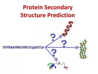
Secondary Protein Structure. What to Know. You will only be tested on what is discussed in class Pay particular attention to topics that are stressed or mentioned several times Most important are the general principles, not details that require memorization

E N D
What to Know You will only be tested on what is discussed in class Pay particular attention to topics that are stressed or mentioned several times Most important are the general principles, not details that require memorization • Know the classes of secondary structure • Types of helices and how amide plane influences secondary structure of helices • Types of pleated sheets • main differences between the structure of an α-helix and a β-pleated sheet • What is the main cause of sterically forbidden regions of ф and Ψ ? • What type bonds stabilize secondary structures? • How do you use the helical wheel? • What influences stability of different helices and the different types of β-pleated sheets?
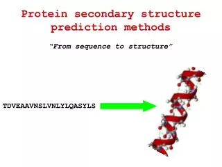
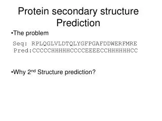
PROTEIN SECONDARY STRUCTURE • Relatively short-range in globular proteins. • Usually long-range in fibrous proteins. • All ф bond angles are equal and all Ψ bond angles are equal providing a repetitive (periodic) structure. • Stability attained through H-bonds.
Main Classes of Secondary Structure All these are local structures that are stabilized by hydrogen bonds • Alpha helix • Beta sheet (composed of "beta strands") • Tight turns (beta turns or beta bends)
What Are the Elements of Secondary Structure in Proteins, and How Are They Formed? • The amide or peptide bond planes are joined by the tetrahedral bonds of the α-carbon. • The rotation parameters are φ and ψ. The conformations shown corresponds to φ= 180° and ψ= 180°. • One can specify a polypeptide’s backbone conformation by the torsion angles (rotation angles) about the Ca-N bond (f) and Ca-C bond (y) of each of its amino acid residues
Consequences of the Amide Plane • Two degrees of freedom per residue for the peptide chain • Angle about the Cα-N bond is denoted φ (phi) • Angle about the Cα-C bond is denoted ψ (psi) • The entire path of the peptide backbone is known if all φ and ψ angles are specified • Some values of φ and ψ are more likely than others.
Many of the possible conformations about an α-carbon between two peptide planes are forbidden because of steric crowding. Several noteworthy examples are shown here.
Protein Structure • Sterically forbidden conformations are those in which any nonbonding interatomic distance is less than its corresponding van der Waals distance • G. N. Ramachandran was the first to demonstrate the convenience of plotting phi,psi combinations from known protein structures • The sterically-favorable combinations are the basis for preferred secondary structures
Steric Constraints on φ & ψ Ramachandran diagram showing the sterically reasonable values of the angles φ & ψ. Shaded regions indicate particularly favorable values of these angles. Dots in purple indicate actual angles measured for 1000 residues (excluding glycine, for which a wider range of angles is permitted) in eight proteins.
Hydrogen Bonds in Proteins A hydrogen bond between a a backbone C=O and a backbone N-H in an acetylcholine binding protein of a snail, Lymnaea stagnalis. Schematic drawing of a hydrogen bond between a backbone C=O and a backbone N-H.
Protein Structure • Helical Structures • if a polypeptide chain is twisted by the same amount about each of its Ca atoms, it assumes a helical conformation • helix characterization • n=number of peptide units per helical turn • pitch=distance helix rises along axis/turn • helixes have chirality
Protein Structure • Helical Structures • If a particular helix is to be more than transient, it must be stabilized (H-bonds) • Only one helical polypeptide conformation has simultaneously allowed conformation angles and a favorable hydrogen binding pattern: a=helix, a particularly rigid arrangement of the polypeptide chain. Designation: 3.613 p=5.4Å
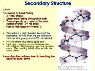
The α-Helix Four different representations of the α-helix First proposed by Linus Pauling and Robert Corey in 1951 A ubiquitous component of proteins, stabilized by H bonds
Protein Structure • Other Helical Structures • 310 helix: p=6.0Å frequently occurs as a single turn transition between the end of an a-helix and the next portion of the polypeptide chain • p helix (4.416 helix): p=5.2Å comparatively wide and flat conformation results in axial hole too small to admit H2O yet too wide to allow van der Waals associations across the helix axis
Exposed N-H and C=O groups at the ends of an α-Helix can be “capped”. Four N-H groups at the N-terminal end of an α-helix and four C=O groups at the C-terminal end lack partners for H-bond formation.
An amphiphilic helixin flavodoxin: A nonpolar helix incitrate synthase: A polar helixin calmodulin:
The Beta-Pleated Sheet • Formed through by side-by-side alignment of polypeptide strands • Strands may be parallel or antiparallel • Stability arises via H-bond interactions • Distance: 3.5Ao for antiparallel strands 3.3Ao for parallel strands • Each strand of a beta sheet may be pictured as a helix with two residues per turn
An antiparallel β-pleated sheet. R groups project alternately above and below the plane of the sheet. Sheet structure is derived from the tetrahedral placement of substituents on the α carbon atoms. This is the more stable form of a β-sheet.
Arrangement of hydrogen bonds in (a) parallel and (b) antiparallel -pleated sheets. Parallel sheets: Large; hydrophobic on both sides of sheet; interior of globular proteins. Antiparallel sheets: 2-3 strands; amphipathic allowing good boundaries with aqueous surroundings.
Protein Structure • Beta structure • b-pleated sheets in globular proteins typically exhibit a right-handed twist when viewed along their polypeptide strand • twists serve important role since b sheets often form central core of proteins • in globular proteins, b sheets are common • parallel b sheets of less than 5 strands are rare, suggesting they are less stable than antiparallel sheets (H-bonds are distorted for parallel sheets)
Protein Structure • Beta structures • mixed parallel-antiparallel sheets are common but occur less frequently than expected from random mixing of strands
The Beta Turn • allows the peptide chain to reverse direction • carbonyl C of one residue is H-bonded to the amide proton of a residue three residues away • proline and glycine are prevalent in beta turns
The structures of two kinds of -turns (also called tight turns or -bends). Proline and glycine are frequently situated in positions 2 and 3, respectively.
Protein Structure • Beta structures • links between 2 antiparallel sheets=hairpin turn • links between 2 parallel sheets=cross-over connection
Fibrous Proteins • Much or most of the polypeptide chain is organized approximately parallel to a single axis • Fibrous proteins are often mechanically strong • Fibrous proteins are usually insoluble • Usually play a structural role in nature
Alpha Keratin • Found in hair, fingernails, claws, horns and beaks • Sequence consists of 311-314 residue alpha helical rod segments capped with non-helical N- and C-termini • Primary structure of helical rods consists of 7-residue repeats: (a-b-c-d-e-f-g)nwhere a and d are nonpolar. Promotes association of helices!
Both type I and type II a-keratin molecules have sequences consisting of long, central rod domains with terminal cap domains. Asterisks denote domains of variable length. • (b) The rod domains form coiled coils consisting of intertwined right-handed a-helices. These coiled coils then wind around each other in a left-handed twist. Keratin filaments = twisted protofibrils (each a bundle of four coiled coils)
Beta Keratin • Found in silk fibers • Alternating sequence:Gly-Ala/Ser-Gly-Ala/Ser.... • Since residues of a beta sheet extend alternately above and below the plane of the sheet, this places all glycines on one side and all alanines and serines on other side! • This allows Glys on one sheet to mesh with Glys on an adjacent sheet (same for Ala/Sers)
Silk fibroin consists of a unique stacked array of b-sheets. The primary structure of fibroin molecules consists of long stretches of alternating glycine and alanine or serine residues. When the sheets stack, the more bulky alanine and serine residues on one side of a sheet interdigitate with similar residues on an adjoining sheet. Glycinehydrogens on the alternating faces interdigitate in a similar manner, but with a smaller intersheet spacing.