Download

1 / 38
680 likes | 2.88k Vues
Pulse Oximetry. Dr. Abdul- Monim Batiha Assistant Professor Critical Care Nursing Philadelphia university . Pulse oximetry is a noninvasive monitoring technique used to estimate the measurement of arterial oxygen saturation (Sao2) of hemoglobin.

E N D
Pulse Oximetry Dr. Abdul-MonimBatiha Assistant Professor Critical Care Nursing Philadelphia university
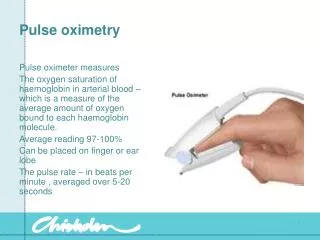
Pulse oximetry is a noninvasive monitoring technique used to estimate the measurement of arterial oxygen saturation (Sao2) of hemoglobin. • Oxygen saturation is an indicator of the percentage of hemoglobin saturated with oxygen at the time of the measurement . • The reading, obtained through pulse oximetry, uses a light sensor containing two sources of light (red and infrared ) that are absorbed by hemoglobin and transmitted through tissues to a photodetector.
The infrared light is absorbed by the oxyhemoglobin ,and the red light is absorbed by the reduced hemoglobin. • The amount and type of light transmitted through the tissue is converted to a digital value representing the percentage of hemoglobin saturated with oxygen. • Oxygen saturation values obtained from pulse oximetry (Spo2) represent one part of a complete assessment of a patient's oxygenation status and are not a substitute for measurement of arterial partial pressure of oxygen (PaO2) or of ventilation (as measured by arterial partial pressure of carbon dioxide(PaCO2)
The accuracy of Spo2 measurements requires consideration of many physiologic variables. Patient variables include the following 1- Hemoglobin level, 2- Arterial blood flow to vascular bed, 3- Temperature of digit or the area where the oximetry sensor is located, 4- Patient oxygenation ability, 5- Fraction of inspired oxygen (percentage of inspired oxygen), 6- Evidence of ventilation perfusion mismatch, 7- Amount of ambient light seen by the sensor, 8- Venous return at the sensor location.
A complete assessment of oxygenation includes evaluation of oxygen content and delivery, which includes the following parameters: • PaO2, • Sao2, • Hemoglobin, • Cardiac output, • Mixed venous oxygen saturation (when available)
Normal oxygen saturation values are 97% to 99% in a healthy individual on room air. • An oxygen saturation value of 95% is clinically accepted in a patient with a normal hemoglobin level. • Using the – oxyhemoglobin dissociation curve, an oxygen saturation value of 90% is generally equated with a PaO2 of 60 mm Hg.
Tissue oxygenation is not reflected by arterial or oxygen saturation obtained by pulse oximetry. • The affinity of hemoglobin to oxygen may impair or enhance oxygen release at the tissue level.
Oxygen is more readily released to the tissues (decreased oxygen affinity) when • Decreased Ph (acidosis), • Increased PaCO2 (respiratory acidosis) • Increased body temperature (hyperthermia) • Increased 2,3-diphosphoglycerate level (a by –product of glucose metabolism also found in stored blood products)
When hemoglobin has greater affinity for oxygen, less is available to the tissue (increased oxygen affinity) increase oxygen binding to the hemoglobin and limit its release to the tissue. Conditions such as • Increased ph (alkalosis) • Decreased PaCO2 (respiratory alkalosis) • Decreased temperature (hypothermia) • Decreased 2,3-diphosphoglycerate level.
Oxygen saturation values may vary with the amount of oxygen usage by the tissues . In some patients, there is a difference in Spo2 values at rest compared with values during activity , such as ambulation or positioning. • Oxygen saturation does not reflect the patient's ability to ventilate. The true measure of ventilation is determination of the PaCO2 in arterial blood .
Use of Spo2 in a patient with obstructive pulmonary disease may result in erroneous clinical assessment of condition. As the degree of lung disease increases, the patient's drive to breathe may shift from an increased carbon dioxide stimulus to a hypoxic stimulus.Enhancing the patient's Spo2 may limit his or her ability to ventilate. • The normal baseline Spo2 for a patient with known severe restrictive disease and more definitive methods of determining the effectiveness of ventilation must be known before considering intervention that enhance oxygenation
Any discoloration of the nail bed can affect the transmission of light through the digit. dark nail polish, such as blue, green, brown, or black colors, and bruising under the nail can limit the transmission of light and result in an artificially decreased Spo2 value. If the nail polish cannot be removed, the sensor can be placed in a lateral side-to-side position on the finger to obtain readings if no other method of sampling the arterial bed is available • Pulse oximetry has not been shown to be affected by the presence of an elevated bilirubin
Pulse oximeters are unable to differentiate between oxygen and carbon monoxide bound to hemoglobin. Readings in the presence of carbon monoxide are falsely elevated. pulse oximetry should never be used in suspected cases of carbon monoxide exposure. An arterial blood gas reading always should be obtained to determine the accurate oxygen saturation. • It has been suggested that dark skin may affect the ability of the pulse oximeter to detect arterial pulsations. one study found a more frequent difference between the spo2 and sao2 with black patients compared with lighter skinned patient ,another study did not find a significant difference
A pulse oximeter should not be used as a predictive indicator of the actual arterial blood gas saturation • A pulse oximeter should never be used during a cardiac arrest situation because of - The extreme limitations of blood flow during cardiopulmonary resuscitation - The pharmacological action of vasoactive agents administered during the resuscitation effort
Equipment • Oxygen saturation meter and monitor, • Oxygen saturation cable and sensor, • Manufacturer's recommended germicidal agent for cleaning the non- disposable sensor (used for cleaning between patients).
Patient assessment • Assess signs and symptoms of decreased ability to oxygenate to determine the need for continuous pulse oximetry monitoring. Anticipation of conditions in which hypoxia could be present allows earlier intervention before unfavorable outcomes occur • Cyanosis • Dyspnea • Tachypnea • Decreased level of consciousness • Increased work of breathing • Loss of protective airway • Agitation • Confusion • Disorientation • Tachycardia • Bradypnea
Assess the extremity (digit) or area where the sensor will be placed to identify factors that may inhibit accuracy of the measurement of oxygenation before attempting to obtain the spo2 reading to enhance the validity of the measurement and allow for correction of factors as possible • Decreased peripheral pulse • Peripheral cyanosis • Decreased body temperature • Decreased blood pressure • Exposure to excessive environmental light source (e.g., examination lights) • Excessive movement or tremor in the digit • Presence of dark nail polish or bruising under the nail • Presence of artificial nails • Clubbing of digit tips
Patient preparation • Explain the need for determination of oxygen saturation with a pulse oximeter to inform the patient of the purpose of monitoring and to enhance patient cooperation and decrease patient anxiety • Explain that the value displayed may vary by patient movement, amount of environmental light, patient level of consciousness ( awake or asleep), and position of the sensor to decrease patient and family anxiety over the constant variability of the values
Explain that the use of pulse oximetry is part of a much larger assessment of oxygenation status to prepare the patient and family for other possible diagnostic tests of oxygenation (e.g., arterial blood gases) • Explain the equipment to the patient to facilitate patient cooperation in maintaining sensor placement
Explain the need for an audible alarm system for determination of oxygen saturation values below a set acceptable limit .Demonstrate the alarm system, alerting the patient and family to the possibility of alarms, including causes of false alarms to provide an understanding of the use of an alarm system and its importance in the overall management of the patient and of circumstances in which a false alarm may occur assists in patient understanding of the values seen while at the bedside.
Explain the need to move or remove the sensor on a routine basis to prevent complications related to the type of sensor used or the degree of tightness in which the sensor is secured around the finger to provide an understanding of the need to move the sensor routinely assists in patient understanding of the frequency of sensor movement. • Ensure that the patient understands pre-procedural teaching. Answer questions as they arise, and reinforce information as needed to evaluate and reinforce understanding of previously taught information.
Procedure • Wash hands to reduce the transmission of microorganisms and body secretions (standard precautions) • Use personal protective equipment to reduce the transmission of microorganisms and body secretions (standard precautions) • Select the appropriate pulse oximeter sensor for the area with the best pulsatile vascular bed to be sampled to obtain accurate spo2 measurements (Use of finger sensors produce the best results over other sites) to optimize signal capture and minimize artifact-related difficulties ( several different types of sensors are available, including disposable and non-disposable sensors that may be applied over a variety of vascular beds)
Do not use one manufactur's sensor with another manufacturer's pulse oimeter unless compatibility has been verified • Select desired sensor site .If using the digit ,assess for warmth and capillary refill. Confirm the presence of an arterial blood flow to the area monitored because adequate arterial pulse strength is necessary for obtaining accurate Spo2 measurements • Avoid sites distal to indwelling arterial; catheters, blood pressure cuffs, military antishock trousers (MAST),or venous engorgement (e.g., arteriovenous fistula, blood transfusions) to obtain accurate Spo2 measurements.
Plug oximeter into grounded wall outlet if the unit is not portable. If the unit is portable , ensure sufficient battery charge by turning it on before using . Plug patient cable into monitor to decrease occurrence of electrical interference (portable systems have rechargeable batteries and depend on sufficient time plugged into an electrical outlet to maintain the proper level of battery charge. When system is used in the portable mode, always check battery capacity.
Check battery capacity always when the system is used in the portable mode ( portable systems have rechargeable batteries and depend on sufficient time plugged into an electrical outlet to maintain the proper level of battery charge)
Apply the sensor in a manner that allows the light source (light- emitting diodes) to be directly opposite the light detector (photodetector) to determine a pulse oximetry value properly,Shielded from excessive environmental light because light from sources such as examination lights or overhead lights can cause elevated oximetry values ( if the oximeter sensor fails to detect a pulse when perfusion seems adequate, excessive environmental light (overhead examination lights,phototherapy lights,infrared warmers) may be blinding the light sensor. Troubleshoot by reapplying the sensor or shielding the sensor with a towel or blanket.
Positioned so that all sensor emitted light comes in contact with perfused tissue beds and is not seen by the other side of the sensor \or without coming in contact with the area to be read because if the light is seen directly from the sensor without coming in contact with the vascular bed , too much light can be seen by the sensor, resulting in either a falsely high reading or no reading ( known as optical shunting, the light bypass the vascular bed; shielding the sensor does not eliminate this if the sensor is too large or not properly positioned
Gently position the sensor so that it does not cause restriction to arterial flow or venous return because the pulse oximeter is unable to distinguish between true arterial pulsations and fluid waves (e.g., venous engorgement or fluid accumulation) • Restriction of arterial blood flow can cause a falsely low value and lead to vascular compromise , causing potential loss of viable tissues. • Edema from restriction of venous return can cause venous pulsation. Evaluating the site above the level of the heart reduces the possibility of venous pulsation (Moving the sensor to another site on a routine schedule also reduces tissue compromise
Never place the sensor on an extremity that has decreased or absent sensation because the patient may not be able to identify discomfort or the signs and symptoms of loss of circulation or tissue compromise • Plug sensor into oximeter patient cable to connect the sensor to the oximeter, allowing Spo2 measurement and analysis of waveforms • Turn instrument on with the power switch • Allow 30 seconds for self-testing procedures and for detection and analysis of waveforms before value are displayed
Determine accuracy of detected waveform by comparing the numeric heart rate value with that of a monitored heart rate or an apical heart rate or both ( if there is insufficient arterial blood flow through the sensor, the heart rate values vary significantly. (consider moving the sensor to another site, such as the earlobe or the nose) ( if the pulse rate detected by oximeter does not correlate with the patient's heart rate, the oximeter is not detecting sufficient arterial blood flow of accurate values ( this problem occurs particularly with the use of the fingers and the toes in conditions of low blood flow .
Set appropriate alarm limits according to the patient's condition. (oxygen saturation limits should be 5% less than patient acceptable baseline & heart rate alarm should be consistent with the cardiac monitoring limits (if monitored) • Wash hands to reduce transmission of microorganisms to other patients • Cleanse non-disposable sensor, if used, between patients with manufacturer's recommended germicidal agent to reduce transmission of microorganisms to other patients
Unexpected outcome 1- Accurate pulse oximetry is not obtainable because of movement artifact. 2- Low perfusion state or excessive edema prevents accurate pulse oximetry measurements. 3- Disagreements occur in Sao2 and oximeter Spo2
Patient monitoring and care • Evaluate laboratory data along with the patient for evidence of poor oxygenation. (Spo2 values are one segment of a complete evaluation of oxygenation and supplemental oxygen therapy. • Data should be integrated into a complete assessment to determine the overall status of the patients. • If Spo2 is used as an indicator of Sao2 ,an arterial blood gas should be done to determine if the values correlate consistently.
Evaluate sensor site every 2 to 4 hours (if a disposable sensor is used )or every 2 hours (if a rigid encased nondisposable sensor is used ).(assessment of the skin and tissues under the sensor identifies skin breakdown or loss of vascular flow, allowing appropriate interventions to be initiated. • Rotate the site of a reusable sensor every 4 hours
Replace a disposable sensor every 24 hours or more frequently if the securing mechanism is compromised or soiled. • Monitor the site for excessive movement ( excessive movement of the sampled site may result in unreliable saturation values. Moving the sensor to a less physically active site reduces motion artifact; using a lightweight sensor also helps . if the digits are used ,ask the patient to rest the hand on a flat or secure surface
Compare and monitor the actual heart rate with the pulse rate value from the oximeter to determine accuracy of values . (The two numeric heart rate values should correlate closely. A difference in heart rate values may indicate excessive movement or a loss of pulsatile flow detection .
Reportable conditions • Inability to maintain oxygen saturation levels as desired, • Change in skin color, • Loss of warmth of tissue unrelated to vasoconstriction, • Loss of blood flow to the digit, • Evidence of skin breakdown due to the sensor, • Change in color of the nail bed indicating compromised circulation at the nail, • Inability to correlate actual heart rate and pulse rate from oximeter.
Documentation • Patient and family education, • Indications for use of pulse oximetry, • Patient's pulse with Spo2 measurements, • Fraction of inspired oxygen delivered (if patient is receiving oxygen), • Patient clinical assessment at the time of the saturation measurement, • Sensor site, • Simultaneous arterial blood gases (if available), • Recent hemoglobin measurement (if available), • Skin assessment at sensor site, • Oximeter alarm settings, • Events precipitating acute desaturation, • Unexpected outcomes, • Nursing interventions.