Download
1 / 14
140 likes | 144 Vues
Explore how nanotechnology is revolutionizing cancer detection and treatment by targeting tumor-specific cells and delivering precision therapy. Learn about the challenges in cancer research and the potential of nanotechnology in overcoming them.

E N D
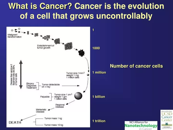
What is Cancer? Cancer is the evolution of a cell that grows uncontrollably 1 1000 1 million 1 billion 1 trillion Number of cancer cells
This evolution results from changes in the cells’ DNA Like a combination lock, many genes must be affected for a cell to become cancer… …but there are many combinations
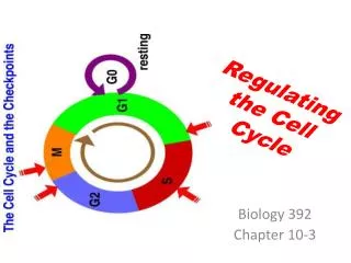
3 major challenges where nanotechnology is needed • Cancer comes from our cells – domestic terrorist! • Cancers are different from patient to patient • Cancers continue to change as they grow
Challenge #1: Cancer comes from our cells – domestic terrorist! Bacteria & viruses = easy to spot Cancer = difficult to detect, difficult to treat
Challenge #2: Cancers are different from patient to patient Each tumor is like a salad from a salad bar They all have a unique combination of ingredients (DNA errors)
Challenge #3: Cancers continue to change as they grow Tumors are playing the lottery, trying to get the right combination to be able to spread (metastasize) Number of cancer cells 1 1000 1 million 1 billion 1 trillion If a tumor is detected too late, it has probably already won
Peptide library on beads Using the body’s own defenses as an early warning system – immune monitoring of tumors Brad Messmer, Thomas Kipps, Dennis Carson Normal serum Cancer serum sort F B red E A D C green Parts of the tumor are oxygen deprived and die
Tumor-specific Uptake of nanoparticles- Roger Tsien Live anesthetized mouse 55 min after tail vein injection Tumor contrast ~6 Cleavable peptide lights up spontaneous mammary tumors in MMTV-polyoma middle T, iNOS -/- mice Minimal cellular binding/uptake protease Maximal cellular binding/uptake Emmi Olson; Dr. Lesley Ellies (UCSD)
Tumor-specific NanoparticleAgregation Nanoparticles are actually much smaller than cells R.Y. Tsien & R. Mattrey (UCSD); P. Daugherty (UCSB) Small nonsticky nanoparticles 20-200 KDa Tumor-specific proteases Mutually adherent aggregates
Nanoparticle smart bombs attack blood vesicles which nourish metastatic tumors -David Cheresh αvβ3-positive αvβ3-negative Tumor Mass
Nanoparticle Smart Bombs for Metastatic Solid Tumors-David Cheresh anb3-Targeting Ligand + + + + + + - + - + + + + - - - - - - - - - - + - Hood et al., Science 2002 + + + + - - - - - - - - - - - - - - - - - - + + + + - + - - - - - - - - - - - - - - - - - - - - + - - + + + + - - - - - - + - - - + - - - - - + + Cationic lipid Nanoparticle targets anb3 and delivers mutant Raf gene to tumor-associated vessels Lung tumor before treatment After treatment 2 related simple drugs against anb3 in Phase II trials (Celengtide –Merck Germany brain cancer) and Phase III trails (Vitaxin MedImmune for melanoma and prostate cancer)
Liposome Encapsulated Virus particles Trogler, Mattrey, Kummel Y Y Y Y Y Y Y Y Y Y • Phase I: Use emulsion and ultrasonic processing of liposomes to construct micron size ultrasonic imaging bubbles. • Phase II: Incorporate adenoviral nanoparticles within nanosized liposome shells for targeting CLL and lymphatic tumors. • Phase III: Incorporate surface receptors for tumor-specific targeting on the liposome mothership surface. Liposome or albumin shell filled with PFC vapor and adenoviral or nanoparticle pay- load with surface targeting receptors Ultrasound image from the Mattrey Lab showing vasculature of lung tumor with PFC filled bubbles. Vascular used to classify tumors
Cancer cell detector for surgical margins in breast cancer • A. Kummel, W. Trogler, I. Schuller, S. Esener, , B. Messmer, D. Messmer, S. Blair, J. Wang-Rodriguez • Cell array to automate touch prep of surgical margins in breast cancer and to detect changes in phenotype with disease progression • Primary detection via size and shape on a-MUC1 coated array • Secondary phenotyping with quantum dot labeled antibodies
Dielectrophoresis (DEP) Separation in Blood Samples – Mike Heller • Top White bacteria are separated from red blood cells by AC electric fields applied to circular electrodes • Bottom 2 types of cells are separated by AC electric field • Can be enhanced with cancer specific nanoparticle markers. • Goal – find rare cancer cells in blood (Nature Biotechnology Vol. 16, 541-546, 1998)