Download
1 / 29
300 likes | 599 Vues
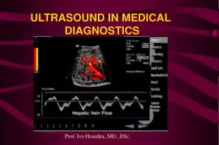
Medical Ultrasound Measurements Lab Tour. Research Highlights. Ultrasound (US) Signal processing Image processing Multiple medical imaging modalities registration Clinically significant evaluative tools Cardiac function Neovascularization Therapeutic ultrasound

E N D
Research Highlights • Ultrasound (US) • Signal processing • Image processing • Multiple medical imaging modalities registration • Clinically significant evaluative tools • Cardiac function • Neovascularization • Therapeutic ultrasound • Acute Cardiac Pacing by High Intensity Focused US
A feature dependent approach for improving speckle noise reduction and side lobes suppression in ultrasound images • Frequency compounding is applied to selected regions in the image and avoided from others, depending on the identification of features for which preserving the axial resolution is of high importance. Such features are point reflectors, lesions boundaries and cysts boundaries. • Apodization is applied only at specific regions in which side lobes are identified.
Reference image Feature dependent compounding Ordinary apodization Ordinary compounding Feature dependent apodization
Automatic detection of left ventricular anatomic features: Mitral valve
מבט מכיוון כפות הרגליים יד שמאל עצם יד שמאל רקמת שומן -בטן ריאה שמאל חדר שמאל חדר ימין שריר הלב ריאה ימין עמוד שדרה יד ימין גב עצם יד ימין
מציאת הגבול הפנימי של חדר שמאל לאורך פעימה שלמה – לצורך מדידות קליניות ו/או התאמה להדמיות אחרות
Registration of Ultrasound and MR Cardiac Images • Given a collection of segmented ultrasound (US) and MR cardiac images, our challenge is to register or align them • There are several factors that make this task difficult: • The heart is constantly in motion • The images are almost certainly not of the same slice • Segmentation of the US images is very difficult due to poorer image quality
Procedure • Find the correspondence between homological points
Procedure Continued • The US image is: rotated, translated, scaled, and finally “warped” to bring the two contours into alignment
Arterial tree reconstruction of neovascularization inside a carotid plaque
Clinical Motivation : The disease Atherosclerosis : Atherosclerosis Progression Plaque Generation IMT Thickening : Plaque’s content Inflammation Calcification : Plaque’s types Unstable Plaque Stable Plaque Plaque rupture Pathological result: Stroke and TIA Clinical result:
Objectives • Develop noninvasive quantitative measures of carotid plaque vulnerability, • using Ultrasound Contrast Agents • Assess the neovascularization phenomena in relationship to inflammation • processes and compare to histological indices Methods • Using Ultrasound IU22 Philips machine • Using ultrasound contrast agents • Developing algorithm for data analysis • Validating the algorithm – Manual / Visual analysis or histology analysis
Results Area of neovascularization Vs. Area of the plaque – comparison to histology Intra-plaque neovascularization paths out-plane vessels Saturation artifacts In-plane vessels Tracking after objects inside the plaque
Tumor Vascularization Imaging - Current research Qualitative estimation of blood flow Dynamic contrast-enhanced ultrasonography (DCE-US) Contrast-Enhanced Gray-Scale Ultrasound Lassau et al. , 2010 control treated Zhou et al. , 2011
3D Structural Quantification Quantification using 3D segmentation and Displaying using surface rendering: Improved resolution using 2D Deconvilution http://www.visualsonics.com/vevo2100 Holzman-Gazit, Goldsher, Kimmel 2006 Michailovich, Adam, 2005
Extracorporeal Cardiac Pacing by High Intensity Focused Ultrasound • Possible Application • Emergency Medicine • Cardiac Arrest • Resynchronization Therapy • Electrode Implantation Site Evaluation
Extracorporeal Cardiac Pacing by High Intensity Focused Ultrasound • HIFU • Multi Element • Dual Phase Sequence • Multiple Frequencies • Accentuated Rarefication • Accentuated Pressure
System Highlights • Subjects • Whole Animals • Isolated Hearts • Monitoring by US Imaging • Timing • Evaluation • ECG • Blood Pressure A. Roytberg, “Study of Efficient Pacing of the Myocardium using External High Intensity Focused Ultrasound,” MSc, Thecnion, 2009.
Thanks • Prof. Dan Adam • Noa Bachner Ph.D. • Yael Petrank Ph.D. • Amit Livneh • Assaf Hoogi • Yossi Tsadok • Avinoam Bar Zion • HananKamis • Liron Shlomo • Ran Marom • Tzvi Goldman
Questions • Thank you.