Download
1 / 33
370 likes | 634 Vues
Derivatives of Germ layers. Dr Rania Gabr. Objectives. By the end of this lecture ,the student should be able to: Explain the results of folding List the derivatives of ectoderm List the derivatives of endoderm List the derivatives of mesoderm. Results of folding.

E N D
Derivatives of Germ layers Dr Rania Gabr
Objectives By the end of this lecture ,the student should be able to: • Explain the results of folding • List the derivatives of ectoderm • List the derivatives of endoderm • List the derivatives of mesoderm
Results of folding 1- Embryo change into cylinderical embryo. 2-Transposition between septum transversum and cardiogenic plate( S.T lies cranial then ventral and lastly caudal).
3- Yolk sac is reduced in size ÷d into: a- intraembryonic ( gut). b- extraembryonic ( atrophies). c- yolk stalk (degenerates). 4- Allantois& connecting stalk become dorsal then caudal then ventral.
After Tail Fold • The connecting stalk (primordium of umbilical cord) is attached to the ventral surface of the embryo. • Allantois (a diverticulum of yolk sac) is partially incorporated into the embryo as a part of hindgut.
5- formation of umbilical cord. 6- The oral membrane was cranially ventral. 7- The cloacal membrane and allantois was caudal ventral.
Results of folding • The amniotic cavity enlarged. • The Yolk sac smaller & divided into (intraembryonic Y.S, Yolk stalk& extra embryonic Y.S). • Allantois& connecting stalk shifted caudally. • S.T Shifted anterior to Cardiogenic plate. • The amniotic cavity more enlarged. • Allantois& connecting stalk shifted ventrally and form the umbilical cord which contains the extra embryonic Y.S and stalk. • S.T Shifted caudal to Cardiogenic plate. • * Placenta will face the umblical cord.
Derivatives of the Ectoderm • Ectoderm is divided into: Surface ectoderm Neuroectoderm
Surface Ectoderm Derivatives • Epidermis of the skin • Hair • Nails • Sweat & Sebaceous glands • Mammary glands • Enamel of the teeth • Lens of eye • Epithelium of sensory organs in the inner ear & nose • Anterior lobe of the pituitary gland
Neuroectoderm • Neural Tube • Neural Crest Cells
Neural Tube Derivatives • Central nervous system (Brain and spinal cord) • Peripheral nervous system • Retina • Sensory epithelia of nose & ear • Pineal gland • Posterior lobe of the pituitary gland
Neural Crest Cells Derivatives • Sensory ganglia of the spinal nerves( dorsal root ganglia) • Sensory ganglia of the cranial nerves • Autonomic ganglia • Meninges (Pia mater & Arachnoid mater) of the brain & spinal cord • Schwann cells: Neurolemmal sheath of peripheral nerves • Satellite cells • Melanoblasts of the skin • Suprarenal medulla (chromaffin cells) • Several skeletal & muscular components in the head (derived from pharyngeal arches)
Derivatives of Endoderm Endoderm gives rise to the epithelial lining of: • Trachea • Bronchi • Lungs Respiratory
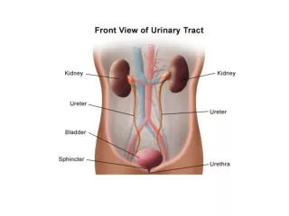
Derivatives of Endoderm Endoderm gives rise to the epithelial lining of: • Gastrointestinal tract • Liver • Pancreas • Urinary bladder • Urachus GIT
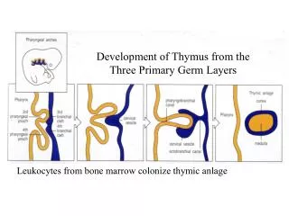
Derivatives of Endoderm Endoderm gives rise to the epithelial lining of: • Pharynx • Thyroid • Tympanic cavity • Pharyngeotympanictube • Tonsils • Parathyroid glands Pharyngeal arches
Derivatives of mesoderm • Connective tissue • Cartilage • Bone • Striated & smooth muscles • Heart • Blood & lymphatic vessels • Kidneys, ovaries, testes& genital ducts • Serous membrane lining the body cavities • Spleen & cortex of the supra renal gland
Development of Somites • As the notochord and neural tube forms • Embryonic mesoderm on each side of them proliferate: • Form thick longitudinal columns of paraxial mesoderm • Each column is continuous with intermediate mesoderm
Development of Somites • Intermediate mesoderm gradually thins into a layer of lateral mesoderm • Lateral mesoderm is continuous with the extraembryonic mesoderm • Extraembryonic mesoderm covers the yolk sac and amnion
Somites • Paraxial mesoderm differentiates and begins to divide into cuboidal bodies called somitesby the end of the 3rdweek • These blocks of mesoderm are located on each side of the developing neural tube
Somites • About 42-44 pairs of somites are present by the end of 5th week • They are triangular in transverse section • Form distinct surface elevations on the embryo • They are used as one of the criteria to know the age of the embryo at this stage
Somites • First appear in the future occipital region • Soon develop craniocaudally • Gives rise to the axial skeleton and associated musculature • Also forms adjacent dermis of the skin • The first pair of somites appear at the end of 3rdweek : day 20
Somites • First appear at a short distance caudal to the cranial end of the notochord • Subsequent pairs form in a craniocaudal sequence
Intraembryonic Coelom • Also known as primordium of embryonic body cavity • Appears as isolated coelomic spaces in the lateral mesoderm and cardiogenic mesoderm • These spaces soon coalesce to form a single horseshoe shaped cavity called intraembryonic coelom
Parietal & Visceral Layers • Somatic or parietal layer continuous with the extraembryonic mesoderm covering the amnion • Splanchnic or visceral layer continuous with the extraembryonic mesoderm covering the yolk sac
Parietal & Visceral Layers • Somatic mesoderm with overlying embryonic ectoderm form the embryonic body wall or somatopleure • Splanchnic mesoderm with underlying embryonic endoderm form the embryonic gut or splanchnopleure
Fate of Intraembryonic Coelom During the 2nd month, the intraembryonic coelom is divided into 3 body cavities: • Pericardial cavity • Pleural cavity • Peritoneal cavity