Download
1 / 63
630 likes | 857 Vues
Basic Techniques to Grow Viruses and Study Virus-Host Interactions. Growth of Viruses. While it is easy to grow bacterial viruses, it is much more difficult and expensive to grow animal viruses Whole animals Embryonating eggs (the classic host for vaccine production).

E N D
Basic Techniques to Grow Viruses and Study Virus-Host Interactions
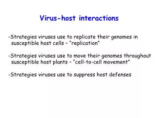
Growth of Viruses • While it is easy to grow bacterial viruses, it is much more difficult and expensive to grow animal viruses • Whole animals • Embryonating eggs (the classic host for vaccine production)
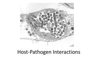
Growth of Viruses, continued • Organ culture - pieces of brain, gut, or trachea, etc. containing different cell types are grown in culture
Organ cultures Sections through tracheal organ cultures: (a) uninfected; (b) infected with a rhinovirus for 36 hours. Note the disorganization of the ciliated cells (uppermost layer) after infection.
Growth of Viruses, continued • Cell or tissue culture – this is where tissues are removed from an organism and are grown “in vitro”, usually in flasks • Primary cultures – are cells that have been directly derived from a tissue and placed in culture. • Are differentiated • They, like the tissue from which they were derived, have a limited life span. • Most will grow attached to the flask as a monolayer of cells one cell thick.
Growth of Viruses, continued • Cell lines • Are dedifferentiated • Are diploid • Survive more passages than primary cell lines, but eventually die • Immortalized cells or continuous cell lines – are cells that have a mutation or mutations that allow the cells to be passaged many times, i.e. they don’t have a limited life span. • Are usually heteroploid • Most were originally derived from a tumor. • Most grow as monolayers, though a few grow in suspension.
Growth of Viruses, continued • When cells grow as monolayers, they can be used to quantify the number of animal viruses using a plaque assay. • The virus is serially diluted in a liquid medium. • For each dilution a set amount is added to a separate plate containing a monolayer of tissue culture cells and the viruses in that solution are allowed to attach to the tissue culture cells. • After attachment has been allowed to occur, a semi-solid medium is added to restrict the movement of new viruses produced so that only adjacent cells will be infected.
Growth of Viruses, continued • Where virus has infected the tissue culture cells, the infected cells will die causing the formation of a clear zone amongst the otherwise intact monolayer of cells • This clear zone is called a plaque and it theoretically represents an area where one virus has infected a single tissue culture cell, has multiplied and been released, and has gone on to infect adjacent cells. • The number of plaque forming units (pfu)/ml can be calculated based on the dilution of the original viral solution. • The term pfu/ml is used rather than the number of viruses/ml because it is possible that occasionally more than one virus infects a single cell. • Often the cells or plaques are stained to help in visualization of the plaques.
Basic Techniques to Study Viruses and Virus-Host Cell Interactions • Serological and immunological methods – these tests are often used for diagnosis of viral infections • May assay directly for the virus (direct assay) • May assay for antibodies, produced in the host, against the virus (indirect assay) • Hemagglutination assay-a direct method to titer virus. • Is based on the ability of some viruses to agglutinate RBCs. • Virus is titered by making serial two-fold dilutions of the virus and determining the highest dilution of virus that causes agglutination of the RBCs.
Serological/Immunological Methods • Hemagglutination-Inhibition Assay – an indirect test for antibody against specific viruses that can agglutinate RBCs. • Mix serial dilutions of patient’s sera with the virus that is the suspected causative agent of the patient’s infection, and then add RBC’s. • If the patient has antibodies specific to the virus, they will bind to the virus and prevent the virus from agglutinating the RBCs.
Serological/Immunological Methods • Immunofluorescence – may be either: • direct and test for the presence of viral antigen in tissues or • indirect and test for the presence of antibodies against a specific virus in a patients sera. • This method uses an antibody with a fluorescent tag attached to it. • With the direct test, the antibody that is tagged is an antibody against the virus that one is testing for. • In the indirect test, the tagged antibody is an antibody against another antibody, i.e. anti-human IgG. The presence of the fluorescent tag is detected by looking under a fluorescent microscope.
Serological/Immunological Methods • ELISA (enzyme linked immunosorbent assay) • Can either be direct (tests for virus) or indirect (tests for antibody to virus). • ELISA is similar to the immunofluorescent assays, but differs in the type of molecule that is tagged to the antibodies that are used. • The molecule that is attached to an antibody in an ELISA assay is an enzyme. • The presence of the enzyme is detected by adding a substrate to the enzyme which when acted upon by the enzyme produces a colored product. • An indirect ELISA test is used to screen individuals for HIV infection.
Direct (sandwich) ELISA (virus?)
Indirect ELISA virus against virus?
Serological/Immunological Methods • Western immunoblot- • A Western immunoblot can be either direct or indirect. • The Western immunoblot analyzes a sample for a specific protein(s) (direct) or for antibodies against a specific protein(s) (indirect). • The screening test to diagnose HIV is the indirect ELISA test. • The indirect Western immunoblot is used to confirm a positive ELISA test.
Basic Techniques to Study Viruses and Virus-Host Cell Interactions • Ultrastructural studies – used for purification purposes • Physical methods • Size by filtration- molecular sieve chromatography. Uses a column filled with beads containing holes. • Large molecules are excluded from the holes and come off the column first. • Small molecules enter the holes in the beads and therefore move slower down the column, coming off the column after large molecules.
Physical methods • Centrifugation • Can pellet materials (virus) by centrifugation • Equilibrium density gradient centrifugation – an inert material is used and it forms a density gradient during the centrifugation. Materials (virus) are forced down until they reach a density that buoys them up. • Rate-zonal centrifugation – similar to density gradient centrifugation, but uses a preformed gradient rather than generating a gradient during the centrifugation process.
Physical methods • Electrophoresis – materials are forced through a meshwork of matrix material (agarose or polyacrylamide) by an electric current. • Usually used for nucleic acids or proteins which are separated on the basis of size, shape, and charge.
Physical methods • Affinity chromatography – Takes advantage of highly specific binding interactions. • A column is made with a material that has a specific receptor (binding interaction) for the substance you are trying to purify (for example the receptor for a particular virus). • A solution from which you wish to purify your virus is run through the column. • The virus binds to the receptor, but everything else is washed through the column. • Next you run a new solution through the column which changes the conditions (pH, ionic strength, etc.) in the column to those in which the specific virus-receptor interaction no longer occurs. • The virus will be eluted from the column.
Physical methods • X-ray crystallography • Chemical methods – to determine the overall composition and the nature of the nucleic acid • Electron microscopy • Whole mounts • + staining (heavy metals) • - staining • Ultrathin sections
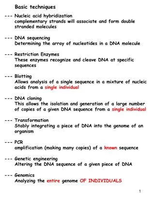
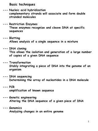
Basic Techniques to Study Viruses and Virus-Host Cell Interactions • Molecular biology – often used to study the structure of the nucleic acid • Hybridization – to come together through complementary base-pairing. • Can be used in identification. • For in situ (or plaque) hybridization the tissue containing the putative organism is treated to release the nucleic acid which is then denatured to single strands. • Labeled single-stranded DNA (a probe) unique to the organism you are testing for is added and hybridization is allowed to occur. • Unbound probe is washed away and the presence of bound probe is determined by the presence of the label.
Molecular Biology • Polymerase chain reaction – used to amplify something found in such small amounts that without PCR it would be undetectable. • Uses two primers, one that binds to one strand of a double-stranded DNA molecule, and the other which binds to the other strand of the DNA molecule, all four nucleotides and a thermostable DNA polymerase. • The primers must be unique to the DNA being amplified and they flank the region of the DNA to be amplified.
PCR • The PCR reaction has three basic steps • Denature – when you denature DNA, you separate it into single strands (SS). • In the PCR reaction, this is accomplished by heating at 950 C for 15 seconds to 1 minute. • The SS DNA generated will serve as templates for DNA synthesis. • Anneal – to anneal is to come together through complementary base-pairing (hybridization). • During this stage in the PCR reaction the primers base-pair with their complementary sequences on the SS template DNA generated in the denaturation step of the reaction.
PCR • The primer concentration is in excess of the template concentration. • The excess primer concentration ensures that the chances of the primers base-pairing with their complementary sequences on the template DNA are higher than that of the complementary SS DNA templates base-pairing back together. • The annealing temperature used should ensure that annealing will occur only with DNA sequences that are completely complementary. WHY? • The annealing temperature depends upon the lengths and sequences of the primers. The longer the primers and the more Gs and Cs in the sequence, the higher the annealing temperature. WHY? • The annealing time is usually 15 seconds to 1 minute.
PCR • Extension – during this stage of the PCR reaction, the DNA polymerase will use dNTPs to synthesize DNA complementary to the template DNA. • To do this DNA polymerase extends the primers that annealed in the annealing step of the reaction. • The temperature used is 720 C since this is the optimum reaction temperature for the thermostable polymerase that is used in PCR. Why is a thermostable polymerase used? • The extension time is usually 15 seconds to 1 minute. • The combination of denaturation, annealing, and extension constitute 1 cycle in a PCR reaction.