Download
1 / 18
200 likes | 559 Vues
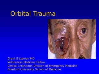
Orbital Imaging. Mounir Bashour, M.D., C.M. What is required when you order orbital MR studies?. 1. Surface coil (orbital or head coil) for better visualization of structures of the orbit 2. Precontrast axial, coronal, and sagittal T1 -weighted images

E N D
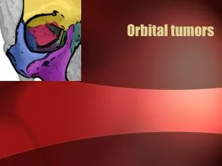
Orbital Imaging Mounir Bashour, M.D., C.M.
What is required when you order orbital MR studies? • 1. Surface coil (orbital or head coil) for better visualization of structures of the orbit • 2. Precontrast axial, coronal, and sagittal T1 -weighted images • 3. Axial, coronal T2-weighted images (fast spin-echo sequences) • 4. Postcontrast axial coronal T1 -weighted images with fat suppression techniques • 5. Sedation in children
What is the strategy in ordering imaging studies in a child with the diagnosis of retinoblastoma? • Diagnosis--Ca++ U/S +/- CT Scan • Extraocular ext. -- MRI orbits • Familial/Bilat --> MRI brain (pinealoblastoma)
What is the strategy in ordering imaging studies in an adult with the diagnosis of intraocular neoplasm? • U/S • IVFA/ICG • MRI--fat suppression
What are paramagnetic agents? • shorten the intrinsic T1 and T2 relaxation times • increase the signal intensity • Melanin, methemoglobin, protein, and gadolinium
In which clinical situations are contrast-enhanced MR studies most helpful in the evaluation of a child with leukocoria? • To differentiate retinoblastoma from Coats' disease.
In which clinical situations are contrast-enhanced MR studies most helpful in the eval-uation of an adult with vitreous hemorrhage? • To differentiate a malignant melanoma of the choroid from a hemorrhagic retinal detachment in age-related macular/extramacular degeneration.
What are the indications for ordering CT orbital studies as a first choice? • 1. Evaluation of orbital trauma • 2. Detection of foreign body • 3. Detection of calcification • 4. Evaluation of osseous, cartilaginous, and fibroosseous lesions • 5. Evaluation of orbital soft tissue lesion with suspicion of bony erosion or detection • 6. Contraindication to MRI
What are the indications for ordering MR orbital studies as a first choice? • 1. Acute proptosis • 2. Suspicion of optic nerve sheath complex lesion • 3. Intraocular tumor with extraocular extension • 4. Detection of wood foreign body • 5. Contraindications to CT
Name the most common orbital lesions showing a well-circumscribed and sharply delineated appearance on CT and MRI in kids • 1. Dermoid cyst • 2. Lymphangioma • 3. Rhabdomyosarcoma • 4. Optic nerve glioma
Name the most common orbital lesions showing a well-circumscribed and sharply delineated appearance on CT and MRI in adults • 1. Cavernous hemangioma • 2. Neurofibroma • 3. Neurilemoma • 4. Fibrous histiocytoma • 5. Lymphoproliferative disorders
Name the most common orbital lesions showing an ill defined appearance on CT andMRI in children • 1. Capillary hemangioma • 2. Idiopathic orbital inflammation • 3. Plexiform neurofibroma • 4. Leukemic infiltrate • 5. Eosinophilic granuloma
Name the most common orbital lesions showing an ill defined appearance on CT andMRI in adults • 1. Idiopathic orbital inflammation • 2. Metastasis • 3. Leukemic infiltrate • 4. Primary malignant tumor • 5. Lymphoproliferative disorders
Which are the ocular and orbital tissues that do not normally enhance on postcontrast MR studies? • 1. Lens • 2. Vitreous • 3. Sclera • 4. Orbital fat • 5. Optic nerve sheath complex
In which clinical situation are contrast-enhanced MR studies most helpful in the evaluation of a patient with proptosis? • Well circumscribed lesions
What are the indications for MR angiography and carotid angiography when imaging orbital lesions? • None
How can you differentiate optic nerve from optic nerve sheath lesions with CT and MR studies? • MRI • Normal nerve does not demonstrate any enhancement • Optic nerve tumor or inflammation demonstrates central enhancement • Optic nerve sheath neoplasm or inflammation demonstrates peripheral enhancement • A cystic or hemorrhagic lesion does not enhance