Download
1 / 37
420 likes | 738 Vues
Pathology Quiz - Liver. Chris Kwan PBL28. Healthy Liver. Smooth capsule Normal size Hepatic vein not obstructed, normal size Even colouration No fibrosis. Fatty liver. Fatty liver. Hepatic Steatosis (Fatty Liver). Two types of fatty liver: Alcoholic steatohepatitis

E N D
Pathology Quiz - Liver Chris Kwan PBL28
Healthy Liver • Smooth capsule • Normal size • Hepatic vein not obstructed, normal size • Even colouration • No fibrosis
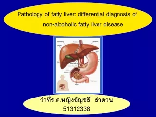
Hepatic Steatosis (Fatty Liver) • Two types of fatty liver: • Alcoholic steatohepatitis • NASH (Non-Alcoholic Steatohepatitis) • Looks paler than the healthy liver • Greasy feel • Lots of lipid droplets amongst hepatocytes • Initially, centrilobular. Eventually entire lobule involved • Normally reversible. However, chronic exposure perivenular fibrosis (not reversible)
Cirrhosis • Cirrhosis = loss of normal liver architecture and presence of hepatocyte nodules • Usual causes of cirrhosis • Alcohol and other drugs • Inflammation (e.g. viruses, autoimmune) • Biliary disorders • Inherited diseases (e.g. haemochromatosis) • Vascular diseases (e.g. thrombosis) • Cryptogenic • An Indian Bit Into Vegetable Curry
Cirrhosis (cont.) • Sometimes can determine cause based on nodule size • Macronodular • Viral hepatitis • Micronodular • Alcohol • Haemochromatosis
Alcoholic Hepatitis • Liver is mottled, normal sized • Microscopic contents: • Mallory Bodies (M) • Neutrophil infiltration (N) • Hepatocyte swelling (H) • Fine subsinusoidal fibrosis (esp. if turning into cirrhosis) • Fatty changes
Mallory Bodies • Consists of hyaline • Has a ropy appearance
Alcoholic Cirrhosis • Resembles viral hepatitis • Shrunken, brown • Microscopic contents: • Parenchymal nodules (N) • Fibrous septaearound nodules (F) • Ischaemic necrosis • Bile stasis • No Mallory Bodies
Haemosiderosis (Haemochromatosis) • Liver looks like milk-chocolate • Caused by deposition of haemosiderin(cellular Fe storage) • Hepatocytes appear brownish too • Excess Fe causes collagen formation • Will see micronodular cirrhosis, which eventually turns into fibrosis • Pancreas also undergoes fibrosis
Acute Viral Hepatitis • Liver is enlarged and red. May turn greenish if cholestasis is present • Ballooning degeneration of hepatocytes • Inflammation • Regenerative changes • Necrosis and apoptosis of hepatocytes
Chronic Viral Hepatitis • Same features as for acute viral hepatitis, PLUS • Lymphoid aggregation + bile duct damage in portal tracts • Steatosis • Interface hepatitis + bridging necrosis fibrosis
Liver infarctions (from thrombosis of hepatic artery and portal vein)
Cardiovascular Diseases • Thrombosis and congestive heart failure can contribute to liver pathology • Thrombosis can ischaemia • Congestive heart failure causes blood to pool in liver makes liver heavy and mottled (nutmeg liver)
Cancer • Variable appearance but it does appear white • Concomittanthaemochromatosis does not colour the cancers • Can appear as a unifocal mass, multifocal nodules or diffusely infiltrative • Can either be well-differentiated or anaplastic • Can metastasise via venous invasion
Primary sclerosingcholangitis (concentric fibrous restructuring of intra- and extrahepatic bile ducts)
Polycystic kidney disease (PKD) that manifested in liver cysts
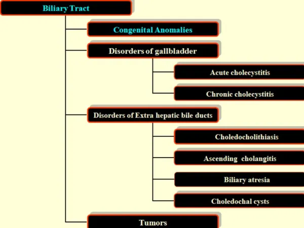
Biliary Disease • Bile duct destruction • Heavy lymphocyte infiltration in portal tracts • Epithelioid cell granulomas also seen • Tends to present with jaundice