Download
1 / 26
300 likes | 682 Vues
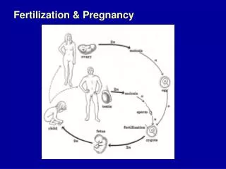
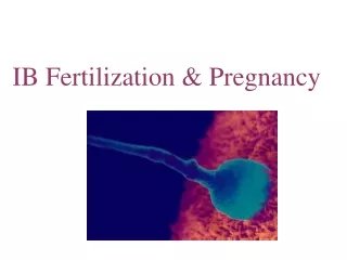
Fertilization, Pregnancy nd Lactation. By Dr. D. Fisher. Fertilization of the Ovum. Takes place in the fallopian tube. Distally, the last 2cm remains spasmatically contracted – under the influence of estrogen for 3 days after ovulation.

E N D
Fertilization, Pregnancy nd Lactation By Dr. D. Fisher
Fertilization of the Ovum • Takes place in the fallopian tube. • Distally, the last 2cm remains spasmatically contracted – under the influence of estrogen for 3 days after ovulation. • Smooth muscle relaxes under the influence of progeterone – secreted by CL. • This allows the fertilized embryo to enter into the uterus.
Implantation of Zygote • Initially the embryo receives its nutrition from the uterus “milk”. • The trophoblastic cells then secrete proteolytic enzymes which digest the stromal cells of the endometrium. • The trophoblastic cells then forms the placenta. • Blood flows from the 16 day after fertilization • 6 weeks after fertilization – placenta takes over the fertilization of the fetus.
Zygote divides to form 2 cells about 18-39 hours after fertilization 2 cells divide to form 4, 8, and so on Pluripotent: Ability to develop into wide range of tissues Morula Solid ball of 12 or more cells Blastocyst or hollow sphere of cells Implantation Burrowing into uterine wall Placenta develops from trophoblast cells Early Cell Division
From conception to birth Three stages Germinal period First 2 weeks of development during formation of primitive germ layers Embryonic period 2nd to end of 8th week, organ systems develop Fetal period Last 30 weeks, organ systems grow and mature Clinical age Mother’s LMP to calculate age of unborn child Postovulatory age Describes timing of developmental events Calculated as 14 days less than clinical age Prenatal Development
The Placenta • Foods cross the placenta by means of diffusion • Permeability of the placenta increases constantly – reaches a peak in the last month – sharp decrease in permeability. Permeability 0 8 20 40
Diffusion of O2 and CO2 • Gradient: Fetus – 30 mmHg Mother - 50 mmHg • Sufficient O2: ???? • Fetal Hb (increase ability to carry O2) • Fetal [Hb] > maternal [Hb] • Bohr effect: increase affinity for O2 in the fetus – decrease affinity in maternal blood. • CO2 gradient: Fetal CO2 – 48 mmHg • Maternal CO2 – 45 mmHg 20 mmHg gradient
Hormonal Control of Pregnancy • Initially the trophoblast cells secrete Human Chorionic Gonadotrophin (HCG) • Functions of HCG: • Prevents the degeneration of the corpus luteum • Stimulates the growth of the CL. • Increase secretion of both estrogen and progesterone • Prevents mestruation
Hormonal Secretion of the Placenta • Corpus luteum required until the 12th week of pregnancy – placenta secretion of hormones takes over completely. • Corpus luteum degenerates. • During pregnancy the placenta secretes: • HCG • Human chorionic somatomammotrophin (HCS) • Estrogen and progestrogen
Functions of HCG • Prevents copus luteum degeneration • Stimulates the interstitial cells of the fetal testes to secrete testosterone • Testosterone responsible for the development of the male sex organs.
Functions of HCS • Starts to be secreted from the 5th week • Increase in HCS secretion is proportional to the weight of the placenta (unterus) • Functions: • Development of the breast • Growth hormone effect enhanced (X200-300) • Decrease maternal insulin sensitivty • Increase [glucose]blood of the fetus • Promotes release of FFA from maternal stores – alternative source of energy for her metabolism.
Functions of Estrogen • Enlargement of the uterus • Enlargement of the breast • Enlargement of the female external genitalia. • Relaxes various pelvic ligaments for easier birth.
Functions of Progesterone • Initially secreted by the Corpus Luteum in moderate quantities • Subsequently, secreted mostly by the placenta. • Action: • Development of the uterine endometrium – essential for the early nutrition of the embryo. • Decrease contractility of the gravid uterus • Contributes to the development of the ovum prior to implantation. • Prepares the breast for lactation.
Fetal Growth • Fetus • At 60 days embryo becomes a fetus • Fetal period • From day 60 to birth is rapid growth • Lanugo • Fine soft hair covering • Vernix caseosa • Waxy coat of protection
Parturition Process by which a baby is born In mother Estrogens overcome inhibitory influence of progesterone Oxytocin is released In fetus Adrenal gland is enlarged prior Labor First stage Onset of regular uterine contraction until cervix dilates to fetal head diameter Second stage From maximum cervical dilation until baby exits vagina Third stage Expulsion of placenta from uterus Parturition
Apgar Scores • Assessment of newborn baby • Appearance, pulse, grimace, activity, respiratory effect • Rated on scale of 0-2, 2 denotes normal function • Total Apgar score is sum from five characteristics
Parturition Physiology • Factors which initiate: • Ratio of estrogens/progesterone increase towards the end of pregnancy • Towards the end of term there is an increase responsiveness of the myometrium to oxytocin. • Increase fetal [oxytocin] towards end of term. • Mechanical stretch of the uterus: • Increase movements of the fetus • Increase size of the fetus • Stretch and/or irritation of cervix uterine – uterine reflex
Lactation: • Function of prolactin: prolactin Promotes the secretion of milk Estrogen and progesterone Birth Decrease [estrogen + progesterone Increase lactogenic effect (prolactin) Increase [milk] in the alveoli of the Breast – not the ducts!!!!!
Ejection of Milk: Oxytocin • Ejection of milk: • Neural reflex • Hormonal reflex spinal cord Suckling of breast Afferent conduction of APs Contraction of the myoepithelial cells Ejection of milk Oxytocin secretion hypothalamus Prolactin secretion Increase [milk] in the alveoli of the breast