Download
1 / 75
800 likes | 1.55k Vues
THE DEVELOPMENT OF B-LYMPHOCYTES. STAGES IN LIFE CYCLE OF B-LYMPHOCYTES. Stage 1 Maturation in bone marrow with development of functional receptors Stage 2 Testing for and elimination of self-reactive receptors Stage 3 Mature naïve cells move to secondary lymphoid tissues Stage 4

E N D
STAGES IN LIFE CYCLE OF B-LYMPHOCYTES • Stage 1 • Maturation in bone marrow with development of functional receptors • Stage 2 • Testing for and elimination of self-reactive receptors • Stage 3 • Mature naïve cells move to secondary lymphoid tissues • Stage 4 • Antigen contact with differentiation into plasma cells and memory cells
B-CELL DEVELOPMENT IN BONE MARROW • Stages are defined by rearrangement and expression of IG genes • Early Pro-B cells • Earliest cells in B-cell lineage • Rearrangement of variable domain of heavy chains • D to J • Late Pro-B cell • Rearrangement of variable domain of heavy chains • V to DJ
B-CELL DEVELOPMENT IN BONE MARROW • Large Pre-B cell • M expressed on cell surface along with surrogate light chains and signal transduction molecules • Pre-B cell receptor • Small Pre-B cell • Pre-B cell receptor not present • Most M chains inside cell • Light chain rearrangement begins
B-CELL DEVELOPMENT IN BONE MARROW • Immature B-cell • Heavy and light chains assembled and transported to surface as IgM receptor complex • Randomness of gene rearrangements leads to self-reactive B-cells • Mature B-cell • IgD expressed on cell surface • Called naïve B-cells
B-CELL DEVELOPMENT IN BONE MARROW • Development depends on non-lymphoid stromal cells • Function of stromal cells • Specific contact through cell adhesion molecules (CAM’s) • VCAM-1 to VLA-4 on early pro-B cells • Produce growth factors for bound B-cells • Stem cell factor (SCF) • Interleukin-7 (IL-7) • Growth factors • Stem cell factor stimulates (G/P) of Early pro-B cells • Interleukin-7 stimulates (G/P) of Late pro-B and L/S pre-B cells
GENE REARRANGEMENTS AND B-CELL SURVIVAL • Gene rearrangement process is imprecise and classified as • Unproductive • Not translated into IG chain • B-cell dies • Productive • Translated into IG chain • Development proceeds • Immunoglobulin loci • Each B-cell has 2 copies on homologous chromosomes • Rearrangements made on both
PROTEINS INVOLVED IN REARRANGEMENT AND EXPRESSION OF IG GENES • Several categories of specialized proteins are required • Lymphoid specific recombination • RAG-1 and RAG-2 • N-nucleotide addition • TdT • Surrogate light chains • Lambda5 and VpreB • Signal transduction • IG-alpha, IG-beta, CD45 and Btk • Transcription factors • EBF and Oct-2
SIGNAL TRANSDUCTION AND BRUTON’S TYROSINE KINASE (BTK) • Bruton’s tyrosine kinase (Btk) • Encoded by gene on X chromosome • Essential for B cell maturation • Mutation in gene • B cells maturation stops at pre-B cell stage • Results in immunodeficiency called • Bruton’s X-linked agammaglobulinemia • Immunodeficiency results in recurrent sinopulmonary infections with • Streptococcus pneumoniae • Haemophilus influenzae
POPULATIONS (SUBSETS) OF B CELLS • B-1 cells (minor subset) • Develop early in embryonic life with unknown origin • Express CD5 (CD5 B cells) • Self-renewing • Primary location is body cavities (pleural / peritoneal) • Produce polyspecific antibodies • B-2 cells (major subset) • Develop after birth • Do not express CD5 glycoprotein • Replaced from bone marrow • Primary location is lymphoid organs • Produce highly specific antibodies
NEGATIVE SELECTION AND FURTHER DEVELOPMENT OF B CELLS • Self reactive immature B cells are either • Eliminated or inactivated • IgM receptor may react with • Cell surface or soluble self antigens • Reactions with cell surface antigens • Induced to commit suicide by apotosis • Clonal deletion • Reactions with soluble antigens • Rendered unresponsive (anergic) to antigen • Maturation continues with reduced surface IgM
SELECTION AND FURTHER DEVELOPMENT OF B CELLS • B cells leave bone marrow and circulate between blood and secondary lymphoid tissues • Secondary lymphoid tissue contains primary lymphoid follicles • Primary lymphoid follicle • Area where B cells congregate in association with specialized stromal cells (follicular dendritic cells) • Passage through primary lymphoid follicle and contact with follicular dendritic cells (FDC) necessary for survival • Few days with no passage • 3 to 8 weeks with passage
CIRCULATION OF B CELLS THROUGH SECONDARY LYMPHOID TISSUES (LYMPH NODES) • Chemokines attract B cells to leave blood and enter cortex of lymph node via high endothelial venule (HEV) • Chemokines attract B cells to lymph node and primary lymphoid follicle • No encounter with antigen • B cells leave node via efferent lymphatic vessel • Anergic B cells detained in T cell area • Induced to commit suicide by apoptosis
B CELL ENCOUNTER WITH ANTIGEN IN SECONDARY LYMPHOID TISSUES • Encounter with antigen takes place in T cell area of lymph node cortex • Antigen reached lymph node from infected tissue via afferent lymphatic vessel • B cell is activated by CD4 T-cell in T-cell area • Activated B cells • Migrate to medulla area and differentiate into plasma cells • Migrate to primary follicle to form germinal center • Migrate to medulla or bone marrow and complete differentiation into plasma cells • Develop into memory B cells
B-LYMPHOCYTE TUMORS • Caused by mutations in genes that regulate cell growth • Genes regulating cell growth • Proto-oncogenes • Promotes cell growth • Tumor suppressor genes • Inhibits cells growth • Mutations in growth regulating genes • Transformation into oncogenes (cancer causing genes)
B LYMPHOCYTE TUMORS • Represents uncontrolled growth of single transformed B cell • Associated with all stages of development • Tumors retain characteristics of cell type and location • Hodgkin’s Lymphoma • Germinal center B cell in lymphoid tissue • Multiple myeloma • Plasma cell in bone marrow • Waldenstrom’s macroglobulinemia • IgM secreting B lymphocyte in lymphoid tissue • Burkitt’s lymphoma • Resembles germinal center B cell in lymphoid tissue
HODGKIN’S DISEASE (LYMPHOMA) • Hodgkin’s disease is a type of lymphoma • Two types of lymphoma • Hodgkin’s disease • Non-Hodgkin’s lymphoma (NHL) • Two main types of Hodgkin’s disease • Classical (95%) • Nodular lymphocyte predominance (5%) • Disease most often starts in lymph nodes of upper body • Chest, neck or under the arms
HODGKIN’S DISEASE (LYMPHOMA) • Cause is not known but there are risk factors • Epstein-Barr Virus (EBV) infection • Geography • United States, Canada, northern Europe • Family history • Identical twin (very high) • Approximately 8,000 new cases each year in US • Cancer cells of HD are unique • Reed-Sternberg cells
WALDENSTROM’S MACROGLOBULINEMIA • Indolent, non-Hodgkin’s lymphoma (NHL) • Classified as • Monoclonal gammopathy • Cancer cells • Features of B-cells and plasma cells • Lymphoplasmacytoid • Located primarily in bone marrow • Produce large amounts of monoclonal protein (antibody) • IgM • Approximately 1,500 new cases each year in US
MULTIPLE MYELOMA • Aggressive, non-Hodgkin lymphoma • Classified as • Monoclonal gammopathy • Cancer cells • Abnormal plasma cells (myeloma cells) • Located primarily in bone marrow • Produce monoclonal proteins (antibody) • IgG, IgA, free kappa or lambda light chains • Approximately 20,000 cases in US for 2008
BURKITT’S LYMPHOMA • B cell tumor with 2 forms • Endemic (African) • Facial tumors • Strongly associated with EBV infection • Nonendemic (Sporadic) • Abdominal tumors • Characteristic translocation • MYC proto-oncogene on chromosome 8 to IG genes • Chromosome 14 (90%) • Chromosomes 2 and 22 (10%) • MYC protein • Normally regulates cell division • Control is lost following translocation to IG gene
CASE STUDY – 21 YEAR OLD MALE • 21 year old WM presents with • Fever (102 F) • Sore throat • Moderate malaise, myalgia and fatigue • Difficulty in swallowing • H and P • Healthy and sexually active • Bilateral anterior and posterior cervical lymphadenopathy • Pharyngeal inflammation • Mild splenomegaly and no jaundice
CASE STUDY – 21 YEAR OLD MALE • Admitted to MC and administered • Penicillin G • Prednisone • Valacyclovir • Laboratory tests • CBC with diff • Liver function tests • Monospot test (Heterophile antibody) • Erythrocyte sedimentation rate (ESR) • Group A streptococcus antigen
CASE STUDY – 21 YEAR OLD MALE • CBC with diff • WBC 12.5 [4.8-10.8] K/uL • RBC 4.0 [3.93-5.22] M/uL • Platelets 250 [150-450] K/uL • Hemoglobin 14.0 [11.2-15.7] g/dL • Hematocrit 40 [34.1-44.9] % • Neutrophils 45 [40-74] % • Lymphocytes 55 [15-47] % • Atypical lymphocytes 18 % • Monocytes 12 [0-12] %
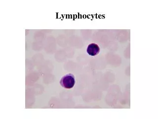
REACTIVE (ATYPICAL) LYMPHOCYTES • Larger in size • Up to 30 um in diameter • More cytoplasm • Less dense nuclear chromatin • Irregular shaped nucleus • Nucleous may be present • Periphery of cell show “scalloped edge”
CASE STUDY – 21 YEAR OLD MALE • Liver function tests • ALT 130 [19-55] U/L • AST 112 [15-37] U/L • Alk phos 150 [50-136] U/L • ESR 40 [0-30] mm/hr • GAS antigen Negative [Negative] • Monospot test Positive [Negative]
CASE STUDY – 21 YEAR OLD MALE • Patient discharged after 36 hours • Treatment • NSAID • Recommendations • Avoid sports • Avoid alcohol