Download

1 / 9
410 likes | 3.94k Vues
Inguinal , femoral and scrotal regions . Anatomy of the inguinal canal . The inguinal canal is approximately 4 cm in length and is located 2 to 4 cm cephalad to the inguinal ligament. In infants, the superficial and deep inguinal rings are almost

E N D
Anatomy of the inguinal canal The inguinal canal is approximately 4 cm in length and is located 2 to 4 cm cephalad to the inguinal ligament. In infants, the superficial and deep inguinal rings are almost superimposed and the obliquity of the canal is slight The canal extends between the internal (deep) inguinal and the external (superficial) inguinal rings. The superficial inguinal ring is a triangular aperture in the aponeurosisof the external oblique muscle and lies 1.25 cm above the pubic tubercle. The deep inguinal ring is a U-shaped condensation of the transversalisfascia and it lies 1.25 cm above the inguinal (Poupart’s) ligament, midway between the symphysis pubis and the anterior superior iliac spine. The anterior boundary comprises mainly the external oblique aponeurosis with the conjoined muscle laterally. The posterior boundary is formed by the fascia transversalis and the conjoined tendon (internal oblique and transversusabdominus medially). The inferior epigastric vessels lie posteriorly and medially to the deep inguinal ring. The superior boundary is formed by the conjoined muscles (internal oblique and transversus) and the inferior boundary is the inguinal ligament.
Content of the inguinal canal • The inguinal canal contains the spermatic cord or the round ligament of the uterus. • The spermatic cord is composed of cremasteric muscle fibers, external and internal spermatic fascia the testicular artery and accompanying veins, the genital branch of the genitofemoral nerve, the vas deferens, the cremasteric vessels, the lymphatics, and the processus vaginalis. • The iliohypogastric and ilioinguinal nerves and the genital branch of the genitofemoral nerve are the important nerves in the groin area • The iliohypogastric and ilioinguinal nerves provide sensation to the skin of the groin, the base of the penis, and the ipsilateral upper medial thigh. • The genital nerve innervates the cremaster muscle and the skin on the lateral side of the scrotum and labia
Covering Peritoneum Transversalis fascia Transversus abdominis m. Internal oblique m. External oblique m. • Obliterated processusvaginalis • Parietal layer of tunica vaginalis • Visceral layer of tunica vaginalis • Internal spermatic fascia • Cremasteric fascia and muscle • External spermatic fascia • Dartos fascia and muscle • Superficialfascia Membranous layer(Scarpa's) Fatty layer (Camper's) • Skin
The Hesselbach triangle The inferior epigastric vessels serve as its superolateral border, the rectus sheath as medial border, and the inguinal ligament as the inferior border. Direct hernias occur within the Hesselbach triangle, whereas indirect inguinal hernias arise lateral to the triangle
Femoral Canal The boundaries of the femoral ring are • • anteriorly by the inguinal ligament; • • posteriorly by Astley Cooper’s (iliopectineal) ligament, the pubic bone and the fascia over the pectineus muscle; • • medially by the concave knife-like edge of Gimbernat’s (lacunar) ligament, which is also prolonged along the iliopectineal line, as Astley Cooper’s ligament; • • laterally by a thin septum separating it from the femoral vein. • A femoral hernia occurs through this space and is medial to the femoral vessels
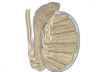
Testis Spermatic cord Ductus deferens Testicular artery Testicular vein (pampiniform plexus) Epididymis:Head,Body,Tail Efferent ductules Rete testis Seminiferous tubule Septum Lobules Visceral layer of tunica vaginalis Cavity of tunica vaginalis Parietal layer of tunica vaginalis Tunica albuginea