Download
1 / 45
500 likes | 888 Vues
The Cranial Nerves. The cranial nerves. 12 in number Are part of the peripheral nervous system All exit the cranial cavity through foramina or fissures All originate from the brain except cranial nerve 11( Accessory nerve) Contain sensory, motor or both components.

E N D
The cranial nerves • 12 in number • Are part of the peripheral nervous system • All exit the cranial cavity through foramina or fissures • All originate from the brain except cranial nerve 11( Accessory nerve) • Contain sensory, motor or both components
-special sensory components are associated with hearing, vision, smelling, balancing and tasting -special motor components include those that innervate muscles derived from the pharyngeal arches
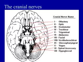
Names of cranial nerves • Ⅰ Olfactory nerve • Ⅱ Optic nerve • Ⅲ Oculomotor nerve • Ⅳ Trochlear nerve • Ⅴ Trigeminal nerve • Ⅵ Abducent nerve • Ⅶ Facial nerve • Ⅷ Vestibulocochlear nerve • Ⅸ Glossopharyngeal nerve • Ⅹ Vagus nerve • Ⅺ Accessory nerve • Ⅻ Hypoglossal nerve
Classification of cranial nerves • Sensory cranial nerves: contain only afferent (sensory) fibers • ⅠOlfactory nerve • ⅡOptic nerve • Ⅷ Vestibulocochlear nerve • Motor cranial nerves: contain only efferent (motor) fibers • Ⅲ Oculomotor nerve • Ⅳ Trochlear nerve • ⅥAbducent nerve • Ⅺ Accessory nerve • Ⅻ Hypoglossal nerve • Mixed nerves: contain both sensory and motor fibers--- • ⅤTrigeminal nerve, • Ⅶ Facial nerve, • ⅨGlossopharyngeal nerve • ⅩVagus nerve
Functional components • General somatic afferent fibers (GSA): transmit exteroceptive and proprioceptive impulses from head and face to somatic sensory nuclei • Special somatic afferent fibers (SSA): transmit sensory impulses from special sense organs of vision, equilibrium and hearing to the brain • General visceral afferent fibers (GVA): transmit interoceptive impulses from the viscera to the visceral sensory nuclei • Special visceral afferent fibers (SVA): transmit sensory impulses from special sense organs of smell and taste to the brain
Functional components • General somatic efferent fibers (GSE): innervate skeletal muscles of eye and tongue • Special visceral efferent fibers (SVE): transmit motor impulses from the brain to skeletal muscles derived from pharyngeal (gill) arches of embryo. These include the muscles of mastication, facial expression and swallowing • General visceral efferent fibers (GVE): transmit motor impulses from the general visceral motor nuclei and relayed in parasympathetic ganglions. The postganglionic fibers supply cardiac muscles,smooth muscles and glands
INTRODUCTION • Oldest sensory modality • 1st Cranial nerve. • Sensory • Detects odor & influence social/sexual behavior.
OLFACTORY EPITHELIUM • Located in upper part of nasal cavity • 3 cell types- • Basal cells • Supporting cell • Olfactory receptor cells • Olfactory receptor cells: Bipolar cells with peripheral and central processes
Unmylinated axons Pass through cribriform plate Enter the olfactory bulb Synapse with mitral cells. OLFACTORY NERVE
OLFACTORY BULB • Ovoid structure possesses several types of nerve cells, the largest is the mitral cell. • Location- inferior to basal frontal lobe. • Highly organized. • Layers : • Glomerular layer • External plexiform layer • Mitral cell layer • Internal plexiform layer • Granular cell layer.
OLFACTORY TRACT • A narrow band of white matter runs from posterior end of olfactory bulb • Projections of Mitral cell axons • Pass posteriorly and divides into medial and lateral striae • Medial stria- • Contralateral olfactory bulb – ant. Commissure. • Lateral stria- carries the axons to the olfactory area of the cortex, prepiriform cortex, periamygdaloid area.
OLFACTORY TRACT & CENTRAL CONECTIONS • Lateral stria- • prepiriform cortex, Iry cortex • Periamygdaloid Nu. • Entorhinal complex (uncus)- IIry cortex
APPLIED ANATOMY • Bilateral anosmia, unilateral anosmia
INTRODUCTION • 2nd cranial nerve • Sensory • Starts from optic disc, extends to optic chiasma • Backwawards continuation of retinal nerve fibers( in the ganglionic layer of retina) • Morphologically & embryologically comparable to sensory tract; it is myelinated and the sheaths are formed by oligodendrocytes
Optic nerve • It leaves the orbital cavity through the optic canal and unites with opposite side to form optic chiasma
INTRAORBITAL PART • Extends from back of eyeball to optic foramina. • Surrounded by the 4 recti muscles near the optic foramina. • Some fibers of superior rectus closely adherent to nerve sheath. • Pierced by central retinal artery/vein.
OPTIC CHIASMA • Situated at the junction of the anterior wall and floor of the third ventricle • Crossroad • Flat & Quadrangular in shape • Partial Decussation
OPTIC TRACTS • Cylindrical bundles of nerve fibers • Ipsilateral temporal fibers & contralateral nasal fibers • Runs outward & backwards • Terminate in the lateral geniculate bodies. • Pupillary reflex fibers pass to superior colliculi.
LATERAL GENICULATE BODIES • Oval structures at posterior termination of optic tract • Consists of six layers of neurons (gray matter) alternating with white matter (formed by optic fibers). • Second order neurons relay station.
OPTIC RADIATION:- • Extends from LGB to Visual Cortex • VISUAL CORTEX:- • Medial aspect of occipital lobe above & below calcarine fissure- • Visual cortex (area 17) • Visual association area (area 18 & area 19)
Oculomotor nerve • Fibers extend from the ventral midbrain, pass through the superior orbital fissure, and go to the extrinsic eye muscles • Components • General somatic efferent fibers (GSE) • General visceral efferent fibers (GVE) • 2 nuclei • Main action-supplies • Superior, inferior and medial recti; inferior obliquus; levator palpebrae superioris • Sphincter pupillea and ciliary muscle • Ciliary ganglion: lies between optic nerve and lateral rectus Oculomotor nerve
Cranial Nerve III: Oculomotor • Functions in raising the eyelid, directing the eyeball, constricting the pupil, and controlling lens shape • The latter 2 functions are parasympathetically controlled • Parasympathetic cell bodies are in the ciliary ganglia
Cranial Nerve IV: Trochlear • Fibers emerge from the dorsal midbrain and enter the orbits via the superior orbital fissures; innervate the superior oblique muscle • Primarily a motor nerve that directs the eyeball • It is the most slender cranial nerve and the only one to leave the posterior surface of brainstem
Cranial Nerve VI: Abducens • Fibers leave the pons and enter the orbit via the superior orbital fissure • Primarily a motor nerve innervating the lateral rectus muscle (abducts the eye; thus the name abducens)