Download

1 / 31
320 likes | 617 Vues
THORACIC CAVITY. LUNGS and PLEURA. Pleura. Visceral pleura: Covers and follows indentations of lung. Parietal pleura: Lines thoracic cavity. Text: p 113, Fig. 1.23. Parietal Pleura Divisions. Costal pleura lines the ribs. Diaphragmatic pleura covers the diaphragm.

E N D
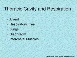
THORACIC CAVITY LUNGS and PLEURA
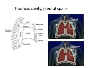
Pleura • Visceral pleura: Covers and follows indentations of lung. • Parietal pleura: Lines thoracic cavity. • Text: p 113, Fig. 1.23
Parietal Pleura Divisions • Costal pleura lines the ribs. • Diaphragmatic pleura covers the diaphragm. • Mediastinal pleura lies against the mediastinum. • Cervical pleura extends above the level of the first rib. • Text: p 115, Fig. 1.24
Pleural Reflections • Costodiaphragmatic recess (space): Space where costal and diaphragmatic pleura meet. • Costomediastinal recess (space): Space where mediastinal and costal pleura meet. • Pulmonary ligament: Transition between visceral and parietal pleura at root of the lung.
Pleural Lines of Reflection • Cervical dome of pleura: Level with neck of the first rib. Anteriorly, 1.5-2.5 cm above the sternal end of the clavicle. Anterior margin extends obliquely behind the sternoclavicular joint. At sternal angle, the pleura is at the median line and two sides stay in contact until the fourth costal cartilage. Text: p 115, Fig. 1.24A
Pleural Lines of Reflection • Right side: • Leaves sternum at 7th costal cartilage. • At 8th costal cartilage at midclavicular line. • At 10th rib at axillary line. • At 11th rib at scapular line. • Extends to level of body of T12 and then ascends.
Pleural Lines of Reflection • Left side: • Leaves sternum at IC space 5. • 1.5 cm from sternal margin at 6th costal cartilage. • Follows same landmarks as right side from this point.
Right Lung Projections • Follows rib 1 to sternoclavicular joint. • Located at median plane at sternal angle. • Extends along median plane from 2nd costal cartilage to 4th costal cartilage. • Leaves sternum at 6th costal cartilage. • At 6th rib at midclavicular line.
Right Lung Projections • At 8th rib at axillary line. • At 10th rib at scapular line. • Ends opposite T11.
Left Lung Projections • Same as right lung except: Cardiac notch begins at 4th costal cartilage. Horizontal at parasternal line. Medial to costochondral junction at 6th cartilage.
Right Lung Morphology • Oblique fissure : Separates superior and inferior lobes. Begins at base of scapular spine. At 5-6th rib at midaxillary line. Ends at 6th costochondral junction. • Text: p 122: Fig. 1.27
Right Lung Morphology • Horizontal fissure : Separates superior and middle lobes. Begins at axillary line. Follows rib 4.
Left Lung Morphology • Oblique fissure: Same as for right lung. • No horizontal fissure.
Lobes of the Lungs • Lobes 3 on right 2 on left Supplied by secondary bronchi
Lobes of the Lungs • Lobules (bronchopulmonary segments) 10 on right 8 on left Supplied by tertiary bronchi • Text: p 125 and 126, Fig. 1.28
Lobes of the Right Lung • Superior lobe: Apical lobule Posterior lobule Anterior lobule • Middle lobe: Lateral lobule Medial lobule
Lobes of the Right Lung • Inferior lobe: Superior lobule Anterior basal lobule Medial basal lobule Lateral basal lobule Posterior basal lobule
Lobes of the Left Lung • Superior lobe: Apicoposterior lobule Anterior lobule Lingular lobule: Homologous to middle lobe of right lung. Superior lobule. Inferior lobule.
Lobes of the Left Lung • Inferior lobe: Superior lobule Anterior basal lobule Medial basal lobule Lateral basal lobule Posterior basal lobule
Bronchopulmonary Tree • Trachea Supported by cartilaginous rings • Primary bronchi Supported by cartilaginous rings • Secondary bronchi Supported by cartilaginous rings • Tertiary bronchi
Bronchopulmonary Tree • Respiratory bronchi • Alveolar sacs • Alveoli
Trachea • Supported by “C”-shaped cartilage segments. • In superior mediastinum. • Anterior to esophagus. • Bifurcates into primary bronchi at sternal angle. • Carina. • Text: p 125, Fig. 1.28
Right Primary Bronchus • Shorter, straighter, and larger than left: Aspirated objects more commonly follow right bronchus. • Gives off three secondary bronchi: Superior to superior lobe of right lung. Middle to middle lobe of right lung. Inferior to inferior lobe of right lung.
Left Primary Bronchus • Twice as long as right primary bronchus. • Gives off two secondary bronchi: Superior to superior lobe of right lung. Inferior to inferior lobe of right lung. • Angle between primary bronchi is 62 degrees.
Bronchopulmonary Tree • Secondary bronchi: Supply lobes. • Tertiary bronchi: Supply lobules (bronchopulmonary segments) • Respiratory bronchi • Alveolar sacs • Alveoli
Lung Vascular Supply • One pulmonary artery to each lung: Part of root of lung: Lobar arteries: One to each lobe Segmental arteries: To segments Intrasegmental
Lung Vascular Supply • Two pulmonary veins from each lung: Intersegmental • Text: p 129, Fig. 1.30
Bronchial Arteries • Supply bronchi • Intrasegmental • Supply tissues of lungs • Two left bronchial arteries from thoracic aorta • Right bronchial artery origin is variable but usually from first aortic intercostal artery. • Text: p 127, Fig. 1.29
Bronchial Veins • Right to azygos vein. • Left to accessory hemiazygos vein or left superior intercostal veins.
Innervation of the Lungs • Anterior and posterior plexus: Lie anterior and posterior to roots of lungs. Contain parasympathetic fibers: From vagus nerve (CN X). Contain sympathetic fibers: From T1-4. Related to esophageal and aortic plexuses posteriorly. Text: p 133, Fig. 1.33