Download
1 / 19
210 likes | 457 Vues
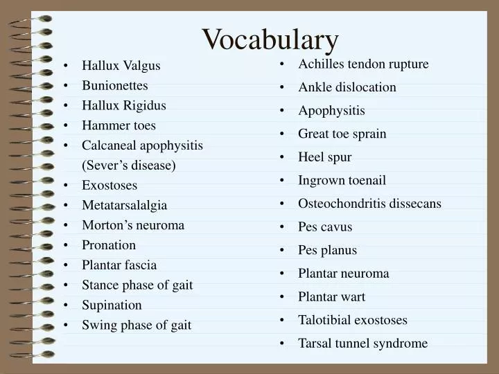
Hallux Valgus Bunionettes Hallux Rigidus Hammer toes Calcaneal apophysitis (Sever’s disease) Exostoses Metatarsalalgia Morton’s neuroma Pronation Plantar fascia Stance phase of gait Supination Swing phase of gait. Achilles tendon rupture Ankle dislocation Apophysitis

E N D
Hallux Valgus Bunionettes Hallux Rigidus Hammer toes Calcaneal apophysitis (Sever’s disease) Exostoses Metatarsalalgia Morton’s neuroma Pronation Plantar fascia Stance phase of gait Supination Swing phase of gait Achilles tendon rupture Ankle dislocation Apophysitis Great toe sprain Heel spur Ingrown toenail Osteochondritis dissecans Pes cavus Pes planus Plantar neuroma Plantar wart Talotibial exostoses Tarsal tunnel syndrome Vocabulary
Arches of the Foot • Why do we have arches? • Supporting body weight • Absorbs shock • Providing space for blood vessels, nerves, muscles • Medial longitudinal arch • Medial border of the calcaneus to distal head of 1st metatarsal • Composed of calcaneus, talus, navicular, 1st cuneiform/metatarsal • Main ligament is the spring ligament and posterior tibialis muscle for reinforcement • Lateral longitudinal arch • Lateral border of the foot • Composed of calcaneus, cuboid,5th metatarsal
Arches of the Foot • Anterior metatarsal arch • Distal heads of the metatarsal • Transverse arch • cuboid and 3rd cuneiform • Plantar fascia (plantar Aponeurosis) • Thick white band of fibrous tissue • From medial tuberosity of calcaneus to proximal heads of metatarsals
Articulations • Interphalangeal articulations • DIP/PIP joints • Produces flexion/extension • Metatarsophalangeal articulations • condyloid type of a joint • Flexion, extension, adduction, abduction • Intermetatarsal articulations • Sliding joints which permits only gliding motions
Articulations • Tarsometatarsal articulations. • Saddle shape allows some gliding and limited flex/ext., Add/abduction. • Intertarsal articulations. • Include the subtalar, midtarsal (transverse tarsal), anterior intertarsal (cuneonavicular). They are sliding joints. • Movements include flex/ext, abduction/add, inversion/eversion, also pronation/supination.
Ligaments of the Foot • Page 405 fig. 17-5 • Subtalar ligaments • Articulation between talus and calcaneus • Talocalcaneal- ant/post, lat/medial • Major ligament is plantar calcaneonavicular= Spring lig. • Midtarsal ligaments • Dorsal talonavicular, bifurcate, dorsal calcaneocuboid • Anterior tarsal joints • Cuneinavicular, cuboideonavicular, intercuneiform, cuneocuboid ligaments. • Dorsal and plantar ligaments
Muscles and Movements • Dorsiflexion/plantar flexion • Plantar- gastrocnemius, soleus, plantaris, peroneous longus, peroneus brevis, and tibialis posterior • Dorsiflexion- -tibialis anterior, extensor digitorum longus, extensor hallucis longus/ brevis, and peroneus tertius muscles • Inversion/adduction/supination • Tibialis posterior, flexor digitorum longus, flexor hallucis longus • Tibialis anterior and extensor hallucis longus • Eversion/Abduction/Pronation • peroneus longus, peroneus brevis • peroneus tertius, extensor digitorum longus • Movement of Phalanges
Anatomy Continued • Nerve supply. • Tibial nerve the largest of the sciatic nerve supplies the muscle of the back of the leg and plantar surface of the foot. • Common peroneal nerve smallest of the sciatic nerve supplies the muscles of the front of the leg and foot. • Blood supply. • The major portion of the blood supplied. • To the foot is by the anterior/posterior tibial arteries.
Foot Biomechanics • Stance Phase or support phase • Starts with initial contact at heel strike and ends at toe off • Foot’s function at heel strike= shock absorber and adapts to uneven surfaces • Heel strike running gait= initial contact of the foot is lat. aspect of calcaneus • In running both feet are off the surface at the same time • Heel strike= leg ext. rotated and foot supinated then the leg int. rotated and foot pronated
Foot Biomechanics • Swing phase or the recovery phase: • Immediately after toe off and the leg is moved from behind the body to the front in preparation for heel strike. • In this phase the leg is external rotated and foot supinates
Evaluation • Structural concerns: • Most people will at some time in their lives develop foot problems • Genetics and habitual use determines your own foot structure • Look for muscular/tendinous tightness, weakness, or hypermobility • Footwear: • Proper footwear (shoe/socks) are essential in injury prevention • Proper shoes for activity • Look for wear on shoes and proper arch support
Evaluation • Surface concerns: • Surfaces that are irregular and vary in resistance can serve to strengthen the foot over time. • A nonyielding surface can lead to acute/chronic injuries • A too resilient/absorbing surface can lead to early fatigue
Assessment • History • How did it happen? Did it happen suddenly or come on slowly? • What was the mechanism? Type of pain? Is there any noises? • Point to the exact site. When is the pain? • What type of surface or footwear are you using ? • Has this ever happened before?
Assessment • Observation • Are they favoring the foot? Are they limping? • Is it deformed, swollen, discolored? • Does it change color by weight bearing or not? • Is the foot well aligned and whether it maintains its shape on weight bearing? • Look for shoe wear.
Medial Medial calcaneus Medial malleolus Talar head Navicular tubercle 1st cuneiform 1st metatarsal 1st MP joint 1st phalanx Lateral Lateral calcaneus Lateral malleolus Peroneal tubercle Cuboid Styloid process of 5th metatarsal 5th metatarsal 5th MP joint 5th phalanx Palpation - Bony Dorsal: 2-4 metatarsal/phalanges, 2-3 cuneiform
Medial/Plantar Tibialis posterior Deltoid ligament Calcaneonavicular lig (spring lig) Medial longitudinal arch Plantar fascia Transverse arch Bursal head of 1st metatarsal Lateral/Dorsal ATF PTF CF Peroneal tendons Extensor tendons of toes Tibialis anterior tendon Palpation – Soft Tissue
Special Tests • Movements and neurological assessment • Extrinsic/instrinsic foot muscles should be assessed for pain & ROM during active, passive, & resistive isometric movement • Tinel test= posterior tibial nerve • Tendon reflex: Achilles tendon (S1 nerve root) • Sensation throughout the whole foot
Special Tests • Pulses: • Taken at the posterior tibial and dorsalis pedis arteries. • Posterior tibial is taken inbetween the medial mallelous and achilles tendon. • Dorsalis pedis is taken inbetween externor hallicus longus and extensor digitorum longus.
Special Test Continued • Flexibility and rigid flatfeet. • Check for flexibility put full weight bearing on foot then lift foot up. • Flexible flatfoot is one that the medial longitudinal arch is flat then an arch is present when weight is removed. • Care is proper shoes, exercise, arch supports or tape.