Download
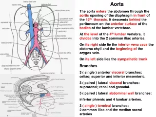
1 / 20
220 likes | 465 Vues
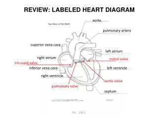
Right common carotid artery. Right subclavian artery. Looking caudal from the aorta. What valve do you see?. ___________ trunk. Aorta/ascending aorta/aortic arch?. Left __________ artery. Right subclavian vein. Left subclavian vein. Superior vena cava (SVC). Left __________ artery.

E N D
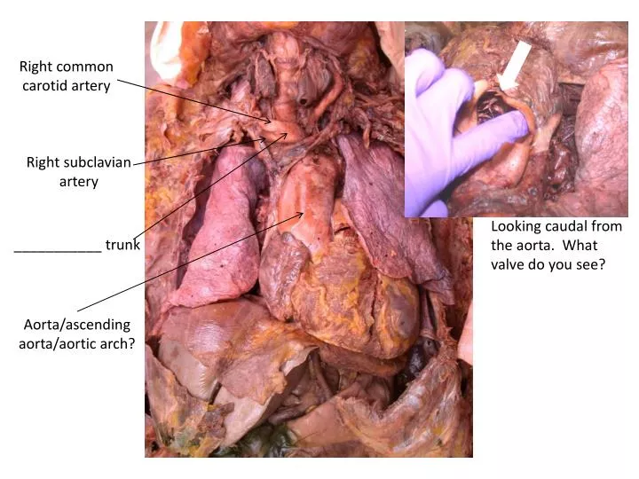
Right common carotid artery Right subclavian artery Looking caudal from the aorta. What valve do you see? ___________ trunk Aorta/ascending aorta/aortic arch?
Left __________ artery Right subclavian vein Left subclavian vein Superior vena cava (SVC) Left __________ artery Right __________ artery
Cranium Right _________ vein Right subclavian vein Left ________ vein Right brachiocephalic vein Superior vena cava (SVC) Abdomen
Left subclavian artery Left axillary artery Medial Lateral What discerns the difference between the subclavian & axillary arteries?
Aorta Right atrium Region of the inferior vena cava Right phrenic nerve Right subclavian vein Superior vena cava Azygos vein
Right atrium Diaphragm Hepatic vein Liver Right ventricle Where is the celiac trunk? Splenic artery Hepatic artery (proper) Common hepatic artery Right gastric artery
Probe indicating hepatic vein (refer to previous image) anatomosis with inferior vena cava (open, with blood clot)
Mark/identify: celiac trunk, splenic artery, common hepatic artery, right gastric artery, hepatic artery (proper) & hepatic vein Cranial Caudal
Cranial Left kidney Mesentery Descending/abdominal aorta Inferior vena cava Inferior mesenteric artery Right kidney Caudal Left common iliac artery Right common iliac artery
Cranial Small intestine / duodenum Superior mesenteric artery Mesentery Right renal artery Left renal vein Inferior vena cava (IVC) Caudal Descending/abdominal aorta
Note the branches of the superior mesenteric artery. The SMA begins to diverge off smaller arteries throughout the mesentery in order to deliver arterial blood to the mesenteric viscera. What organs are serviced by the superior mesenteric artery? Where does this arterial blood drain?
ID all 4 (actually 5) arteries that branch from the dorsal/abdominal/descending aorta that you can see in this image (other than the common iliac arteries). What organs do they deliver arterial blood to? How do those respective organs return venous blood to the heart?
Left renal vein IVC Right renal artery Left ovarian/testicular vein Left ureter
Descending / abdominal aorta Left ______________________artery Inferior mesenteric artery Right exterior iliac artery Right ______________artery _____________________ vein Right common iliac vein Right ovarian artery
Despite clearly visualizing the abdominal vasculature, remember that these arteries and veins are retroperitoneal. Note the progression of images. In order to visualize these major arteries in a patient, you’d have to make an incision in the peritoneum. Abdominal cavity Incised peritoneal membrane to visualize WHICH ARTERIES? Abdominal cavity: bowel reflected cranially
Right external iliac artery “Right inguinal ligament” (where right external iliac artery transitions to right femoral artery) _______ _______ Nerve Right femoral artery
Femoral artery Femoral artery Deep femoral artery
Mesentery Mesentery Notice how mesentric artery on the right is much smaller in diameter (NOT the superior mesenteric artery as it branches off the dorsal/descending/abdominal aorta) within the mesentery. Also note how the probe in the left image is retracting a region of the superior mesenteric vein that will eventually anastomize with the hepatic portal vein.
Trachea Mandible Trachea Mandible Clavicle Clavicle Right common carotid artery Right ________________ vein
Left cephalic vein Left __________________________ Muscle Left median cubital vein Left basilic vein