Download

1 / 30
340 likes | 588 Vues
Medical Biochemistry Molecular Principles of Structural Organization of Cells. 5. NUCLEIC ACIDS. COMPONENTS OF THE NUCLEIC ACIDS. 1. NITROGENOUS BASES The nitrogenous bases are divided into two groups: 1. Purine bases: Purine Adenine (A, Ade) Guanine (G, Gua)

E N D
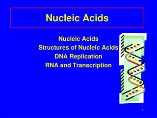
Medical Biochemistry Molecular Principles of Structural Organization of Cells 5. NUCLEIC ACIDS
COMPONENTS OF THE NUCLEIC ACIDS 1. NITROGENOUS BASES The nitrogenous bases are divided into two groups: 1. Purine bases: Purine Adenine (A, Ade) Guanine (G, Gua) 6-aminopurine 2-amino-6-oxopurine 2. Pyrimidine bases Pyrimidine Uracil (U, Ura) Thymine (T, Thy) Cytosine (C, Cyt) 2,4-dioxypyrimidine 5-methyluracil 2-oxo-4-aminopyrimidine
Nitrogenous bases (NB) are classified in: Major bases • Major purine bases are • Adenine (A) • was isolated from pancreas and yeast • in both DNA and RNA, in nucleosides mono-, di-, triphosphates, coenzymes • Guanine (G) • isolated from guano • in both DNA and RNA, nucleosides mono-, di-, triphosphates • Major pyrimidine bases are • Uracil (U) • in RNA, nucleotides, activates the substrates (UDP-glucose), free • Thymine (T) • isolated from thymus DNA • in DNA, and in small amounts in RNA • Cytosine (C) • in both DNA and RNA, nucleosides (cytidin-phosphates) acting in the synthesis of phospholipids Minor bases are primarily found in tRNA and in trace in rRNA; e.g.: 2-methyladenine, 1-methylguanine, 5-methylcytosine, 5-oxymethylcytosine
General properties of the nitrogenous bases • They are hetero-cycles; due to the presence of N have an alkaline character • When H is changed with –OH or –NH2 the solubility in water is reduced, the melting point increases • The bases have the ability to undergo a lactam-lactim (keto-enol) tautomerism • The amine compounds have an alkaline character, the enol compounds act as acids. • At pH<9 (in biological systems) the lactam (keto) form is predominant favoring the formation of covalent bonds of N-glycoside type between N atom in position 1 of pyrimidine or N atom in position 9 of purine and semiacetal –OH (C-1) of pentose • They have a maximum absorbance at =260nm (UV) – used to dose with spectrophotometric method in UV • Low solubility in cold water; soluble in alkaline solutions
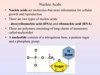
2. PENTOSES -Ribose (R)-2-deoxyribose (dR) in RNA in DNA 3. PHOSPHORIC ACID MOIETY H3PO4- PO3H2 • is able to link the nucleotides forming phosphodiester bond between -OH in position 3’ of the pentose in one nucleotide and -OH in position 5’ in the other nucleotide
NUCLEOSIDES Compounds containing nitrogenous base linked to pentose = nucleosides (C-1’ of pentose is linked to N-9 in purine or N-1 in pyrimidine = N--glycosidic bond) Ribonucleosides R+ A = adenosine R+ G = guanosine R+ C = cytidine R+ U = uridine Deoxyribonucleosides dR+A=deoxyadenosine dR+G=deoxyguanosine dR+C=deoxycytidine dR+T=deoxythymidine
NUCLEOSIDES • Are considered products of the partial hydrolysis of nucleotides • Ribonucleosides exist free in small amounts; deoxyribonucleosides do not exist free Minor nucleosides contain minor nitrogenous bases • exist in tRNA • the most widespread are dihydrouridine, pseudouridine, ribothymidine
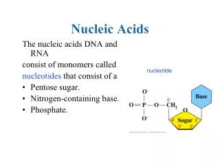
NUCLEOTIDES • They are the monomer units of the nucleic acids; they result from the partial hydrolysis of the nucleic acids under the action of nucleases • They are phosphoric esters of the nucleosides • (nucleotide = nucleoside + H3PO4 = nitrogenous base + pentose + H3PO4) • The phosphate group can add to positions 2’, 3’, 5’ of ribose 3’, 5’ of deoxyribose • adenosine-3’-monophosphate adenosine-5’-monophosphate • Free nucleotides are nucleosides-5’-P (mononucleotides) that are involved in the synthesis of nucleic acids and are formed by their decomposition
Ribomononucleotides R+A+H3PO4 = adenosine-5’-monophosphate = AMP = adenylic acid R+G+H3PO4 = guanosine-5’-monophosphate = GMP = guanylic acid R+C+H3PO4 = cytidine-5’-monophosphate = CMP = cytidylic acid R+U+H3PO4 = uridine-5’-monophosphate = UMP = uridylic acid Deoxyribomononucleotides dR+A+H3PO4 = deoxyadenosine-5’-monophosphate = dAMP = deoxyadenylic acid dR+G+H3PO4 = deoxyguanosine-5’-monophosphate = dGMP = deoxyguanylic acid dR+C+H3PO4 = deoxycytidine-5’-monophosphate = dCMP = deoxycytidylic acid dR+T+H3PO4 = deoxythymidine-5’-monophosphate = dTMP = deoxythymidylic acid
Nucleosidepolyphosphatesare formed by linking an additional phosphate group. • The nucleotides may contain • 1 phosphoric acid moiety - mononucleotides (monophosphate nucleosides), • 2 phosphoric acid moieties - dinucleotides (diphosphate nucleosides), • 3 phosphoric acid moieties - trinucleotides (triphosphate nucleosides), Nucleoside diphosphates and triphosphates are the most frequently occuring in the cells. In the cell, all the nucleoside phosphates occur as anions:AMP2-, ADP3-, ATP3- ADP and ATP are rich in energy = macroergic, used by the organism for performing different functions. Other nucleotides are implicated in the function of biological synthesis.
Ribonucleoside phosphates adenosine-5’-mono-, di-, tri-phosphate = AMP, ADP, ATP guanosine-5’-mono-, di-, tri-phosphate = GMP, GDP, GTP cytidine -5’-mono-, di-, tri-phosphate = CMP, CDP, CTP uridine -5’- mono-,di-, tri-phosphate = UMP, UDP, UTP AMP ADP ATP Deoxyribonucleosides phosphates deoxyadenosine-5’- mono-, di-, tri-phosphate = dAMP, dADP, dATP deoxyguanosine-5’- mono-, di-, tri-phosphate = dGMP, dGDP, dGTP deoxycytidine -5’- mono-, di-, tri-phosphate = dCMP, dCDP, dCTP deoxythymidine-5’- mono-, di-, tri-phosphate = dTMP, dTDP, dTTP
NUCLEOTIDE DERIVATIVES Cyclic nucleotides (3’,5’-AMPc 3’,5’-GMPc) are universal regulators of intracellular metabolism. • cAMP • is mediator of the action of hormones as second messenger, • activates and regulates the function of enzymes – allosteric mechanism in metabolic systems. • cGMP • Second messenger for the action of hormones cAMP cGMP
NUCLEOTIDE DERIVATIVES Nucleotide coenzymes (uridine, cytidine, deoxythymidine, adenosine, guanosine coenzymes) contain residues of glucides, alcohols, aminoacids, lipids, inorganic compounds: • UDP-glucose (UDPGlc, UDPG) is intermediate in the reversible conversion of glucose in galactose, formation of glycogen in animals or starch in plants. • CDP-choline is involved in the formation of phosphatidyl-choline and choline plasmalogens • CMP-sialic acid • UDP-glucuronic acid is a donor of glucuronic acid radical for the coupling reactions of native or foreign substances UDP-G CDP-choline
GENERAL PROPERTIES AND BIOCHEMICAL ROLE OF NUCLEOTIDES • Properties: • Have an acidic character (the protons in the phosphoric acid moiety dissociate: nucleozid-O-PO32-) • Maximum absorbance at =260nm (UV) due to the presence of nitrogenous bases • Nucleotides can be hydrolyzed by 5’-nucleotidase, setting the H3PO4 free • Biochemical role: • In the structure of coenzymes (NAD+, FAD, CoA-SH) • Coenzymes: UDP-G, CDP-Choline • Take part in the enzyme catalyzed reactions: • CTP biosynthesis of phospholipids • UTP in biosynthesis and conversion of carbohydrates • Trinucleotides are precursors in the biosynthesis of nucleic acids • Second messengers for the hormonal control (3’5’-AMPc, 3’5’-GMPc) • ATP is the universal macroergic compound of living organisms
Role and biochemical importance of ATP • ATP, ADP, AMP take part in processes of storage and utilization of the energy set free during the cellular metabolism • They act as donors or acceptors of phosphate moiety • The reaction: ATP-ase ATP + H2O ADP + H3PO4 reflects the energy flow in the cell; it provides the transfer of the chemical energy used in the cellular metabolism. This process implies 2 fundamental aspects: • Formation of ATP represents the storage of chemical energy resulted from the food • Transformation of ATP in ADP represents the generation and use of energy stored in the ATP molecule ATP ADP H3PO4
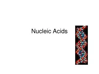
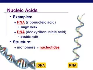
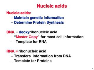
POLYNUCLEOTIDES = NUCLEIC ACIDS • Are macromolecular substances result of the condensation of a great number of mononucleotides (structural units) • They are: • Polyribonucleotides = Ribonucleic acid (RNA) • Polydeoxyribonucleotides = Deoxyribonucleic acid (DNA) • Distinct characters: DNARNA NB: A, G, C, T A, G, C, U Pentose:dRR Number of nucleotide monomers DNA > RNA Length of chain DNA > (except some viruses) Structure double helix 1 chain • Due to the acidic character, nucleic acids are linked with basic proteins, (histones and protamines) and neutal proteins forming deoxyribo-nucleoproteinsribonucleoprotein
STRUCTURE AND LEVELS OF ORGANIZATION OF NUCLEIC ACIDS PRIMARY STRUCTURE DNA and RNA are linear polynucleotide chain made up of mononucleotides linked by 3’,5’-phosphodiester bonds: each pentose 3’-OH of one mononucleotide is linked covalently to pentose 5’-OH of the neighboring mononucleotide. The chains have 2 ends: 5’ endwith triphosphate and 3’ end with a free –OH The chains are polar and directed 5’ 3’ or 3’ 5’ (exception: the circular DNA and RNA of certain viruses and bacteria).
STRUCTURE AND LEVELS OF ORGANIZATION OF NUCLEIC ACIDS PRIMARY STRUCTURE The genetic text of DNA is composed of code triplets or codons = linear sequences of three adjacent nucleotides The sites of DNA chain that contains information on the primary structure of all types of RNA are structural genes. The order of nucleotides in RNA is the same as that in the DNA region that is replicated (copied) with the distinction that RNA consists of ribonucleotides that contain U instead of T
G 3’.5’-phosphodiester bond 3’.5’-phosphodiester bond C A 3’.5’-phosphodiester bond 3’.5’-phosphodiester bond T 3’.5’-phosphodiester bond 3’.5’-phosphodiester bond Primary structure of DNA Primary structure of RNA
SECONDARY STRUCTURE • In 1953 Watson and Crick proposed a double-helix model for the DNA secondary structure • The chains are directed antiparallelly (one chain runs in 5’ 3’ direction and the second 3’ 5’ direction) • The pentose phosphate moieties are directed outwards • The bases protrude into the interior of the helix
Formed by specific pairing of a base of one polynucleotide chain with a base of the other chain. The correspondence of the base pairs is called complementarity • The interaction of A and T is effected through the involvement of 2 H bonds • The interaction of G and C is effected through the involvement of 3 H bonds A = T G ≡ C
SECONDARY STRUCTURE OF DNA Relationship concerning the content of individual bases in DNA (Chargaff, 1949): • A+G = C+T or (A+G)/(C+T) = 1 • A = T or A/T = 1 • G = C or G/C =1 • A+C = G+T or 6-amino group = 6-keto group • (A+T) and (G+C) are the only variable; if: • (A+T)>(G+C) the DNA is AT type • (G+C)>(A+T) the DNA is GC type These rules indicate that the buildup of DNA is effected in a strict conformity with the pairwise interactions A-T and G-C
TERTIARY STRUCTURE OF DNA • The double helical molecule is twisted looking like a supercoil or a bent double-helix • It has a great flexibility; the conformation is not rigid. • There are differences between the native DNA, ”in vivo”, and the one “in vitro”; by removing the water and dependent on the electrolytes in the environment, the double-helix is structurally altered.
TYPES AND LOCATION OF DNA • Nuclear DNA (97-98%) in the chromosomes coupled with basic proteins (protamines, histones) forming chromatine. Nucleolus contains associated DNA and RNA • Mitochondrial DNA (1-3%) in the mitochondria matrix • Structure of simple or double circular helix; does not form complex with proteins; MW << nuclear DNA • Function: • Takes part in maintenance of the mitochondria structure • Contains the information necessary to synthesize specific proteins intra and extra mitochondria • May control the synthesis of the ribosomes • Site of the genetic mutations
GENERAL PROPERTIES OF DNA • COLLOIDAL BEHAVIOR • On dissolution, nucleic acids become swollen and form viscous, colloid-like solutions; the hydrophilicity is mainly determined by the occurrence of phosphate moieties; in solution the nucleic acids exist as polyanions with acidic properties. Double-stranded nucleic acids are less soluble than single-stranded ones • DENATURATION - RENATURATION • Is produced by heating and the action of chemical agents which break hydrogen and van der Waals bonds stabilizing the secondary and tertiary structures. E.g.: heating DNA results in a separation of its double helix (“helix-coil” transition); • Slowly cooled, the chains reunite according to the complementarity principle, DNA regaining its native double–helix; this phenomenon is called renaturation
The helical structure rotate the plane of polarized light exhibiting an optical activity while the breakdown of the spatial arrangement reduce the optical activity to zero. • The DNA absorbs the UV light maximally at 260nm. The absorption intensity of a native nucleic acid is • increased as the DNA is denaturated (hyperchromic effect) or • decreased when the double-helix is reformed (hypochromic effect) • HYBRIDIZATION: • the process whereby hybrid duplexes of complementary DNA and RNA combined. • the aptitude of nucleic acid to renaturate after denaturation has provided a valuable method of cloning different genes and other DNA sequences from different organisms
BIOLOGICAL FUNCTIONS OF DNA • The molecular basis of the transmission of genetic information from one generation to another • Ensures and controls the synthesis of the proteins (enzymes) • In DNA there is encoded the genetic program of development, maintenance and reproduction of each organism • Ensures the differentiation and regulation of cells and the constance of the cell replication • Is the molecular basis of the natural or induced genetic mutations
STRUCTURE AND LEVELS OF ORGANIZATION OF RNASECONDARY AND TERTIARY STRUCTURE Messenger RNA = mRNA • Formed in the cell from pro-mRNA that contains the transcripts of DNA • The code element of mRNA is a linear sequence of three adjacent nucleotides = codon or code triplet. Each codon corresponds to a defined aminoacid. • The secondary structure of mRNA is a bent chain (hairpins and linear regions) • The tertiary structure is like a thread wound round a spool (a special transport protein - informofer)
Transfer RNA = tRNA • The secondary structure of tRNA is a shape of clover-leaf determined by intrachain pairing of complementary nucleotides in certain regions of the chain: • Acceptor region (end or terminus) - 4 linearly linked nucleotides of which CCA sequence is common in all types of tRNA. The 3’ –OH of adenosine is free. At this site the -COOH of the aminoacid is added to be transported to the ribosomes, to be used in the protein synthesis. • Anticodon loop (7 nucleotides) contains a triplet specific for each tRNA = anticodon, complementarily paired to a codon of mRNA; the interaction betweencodon and anti-codon determines the order of the aminoacids in the polypeptide chain • Thymine-pseudouracil (TΨC) loop (7 nucleotides) involved in binding the tRNA to the ribosome • Dihydrouridine loop (diHU) (8-12 nucleotides) binding aminoacyl-t-RNA synthetase, the enzyme which recognizes the aminoacid • Extra loop varies in shape and composition in various tRNA • The tertiary structure – shape of a bent elbow; the cloverleaf loops are folded back on the molecular framework and held together by Van der Waals bonds
Ribosomal RNA = rRNA • Enters in the structure of the ribosomes. n ribosomes + 1 mRNA = polisome • Secondary structure: helical regions alternating with nonhelical bent regions • Tertiary structure constitutes the framework for the ribosome; ribosomes proteins adhere to the tertiary structure on the outside. Chromosomal RNA in nucleus –recognition and activation of DNA genes Low-molecular RNA in nucleus and cytoplasmic RNA particles – activation of DNA genes formation of the skeleton for protein particles involved in the transfer of rNA from nucleus into the cytoplasm