Download
1 / 50
661 likes | 1.31k Vues
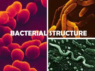
Bacterial Cell Structure. Dr. Zaheer Ahmed Chaudhary Associate Professor Microbiology Department of Pathology. Classification. Based on shape, arrangement and size. Shape : Cocci (round) Bacilli (rod) Spirochets (spiral shape) Pleomorphic (variable shape)

E N D
Bacterial Cell Structure Dr. Zaheer Ahmed Chaudhary Associate Professor Microbiology Department of Pathology
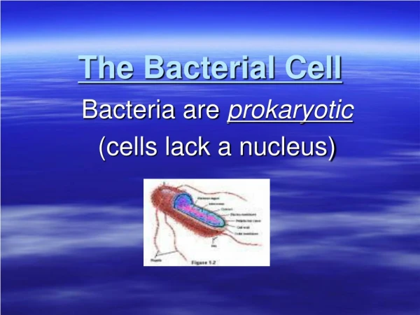
Classification Based on shape, arrangement and size. • Shape : • Cocci (round) • Bacilli (rod) • Spirochets (spiral shape) • Pleomorphic (variable shape) • Shape is determined by rigid cell wall. • Microscopy remains the main stay in identification.
Arrangements: • Diplococci are in short chains (streptococci). • Grape like clusters (staphylococci). • Arrangements depend upon orientation and degree of attachment of the bacteria at the time of cell division.
Size: • Bacteria range in size from mycoplasma, the smallest bacteria (0.2um), to bacillus anthracis, one of the largest bacteria. • Viruses range from polio virus (the smallest virus) to pox virus (the largest virus). • Yeast are larger than bacteria. • Size of bacteria varies from 0.2 to 5um. • Largest bacterial rods are of the same size as of yeast.
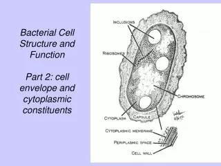
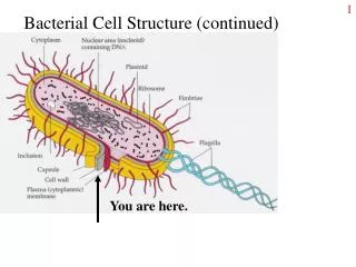
Structure of Cell Wall • Some bacteria have surface features, external to cell wall like capsule, flagella and pili. • Cell wall is a multi-layered structure, located external to cell membrane. • It is composed of inner layer, peptidoglycan and outer layer which is of variable thickness. • Peptidoglycan gives structural support and maintains specific shape of the cell.
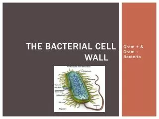
Gram+ & Gram- Cell Wall Composition • Structure, chemical composition and thickness of cell wall varies in G+ and G – bacteria. • Peptidoglycan layer is much thicker in G+ than G- bacteria. • Some G+ bacteria have fibers of teichoic acid, whereas G- bacteria lack it. • In contrast, G- have complex outer layer of lipopolysachride, lipoprotein and phospholipids.
There is a periplasmic space between outer cell wall and cytoplasmic membrane. • It is the site of betalactmase enzyme which degrades penicillin and betalactam drugs.
Properties of Cell Wall • G- bacteria contain endotoxins (lipopolysachrides). • Polysachrides and proteins are antigens. • Porin proteins play role in facilitating the passage of small hydrophillic molecules into the cell. They also act as channels to allow the essential substances like sugar, aminoacids, vitamins, metals and drugs into the cell.
Cell Wall of Acid fast bacteria • Mycobacterium tuberculosis have unusual cell wall which cannot be stained by gram stain. • Bacteria resist decolorization with alcohol after staining with carbolfuchsion. This due to high contents of mycolic acid in the cell wall.
Peptidoglycan • It is complex, interwoven network which surrounds the entire cell. • Composed of single covalently linked macro molecule present only in bacteria to give rigid support to the cell. • It allows the cell to withstand media of low osmotic pressure e.g water. • It consists of peptide and sugar (glycon) which make the molecule.
Carbohydrate is the backbone, which is composed of alternate N acetylemuramic acid and N acetyleglusomin molecule. • Each muramic acid molecule is attached to tetrapeptide consisting of both D-L amino acids, composition of which differs from one bacteria to other bacteria.
Two important aminoacids, diaminopimelic acid and D-alanine which is involved in cross linking of tetra peptide. • Peptidoglycan is not present in human cells. It is a good target for antimicrobials. • Drugs like penicillin, cephalosporin and vancomycin inhibit the synthesis of peptidoglycan by preventing the transpeptidaseengymefrom creating cross linkage between two adjacent tetrapeptides.
Lysozyme • Lysozyme is an enzyme present in human tears, saliva and mucous which can decompose the peptidoglycan backbone by breaking its glycerol bonds and increasing natural resistance of host against bacteria. • The cell swells and ruptures as a result of water entry into the cell after treatment by lysozymes.
Lipopolysachrides (LPS) • LPS of outer membrane of cell wall of G- bacteria is endotoxin, responsible for disease features e.g fever, shock and hypotension. • Endotoxin is the integral part of G- bacteria cell wall. • LPS is composed of 3 distinct units: • phospholipids called Lipid A, responsible for toxic effects. • A core of polysachride of 5 sugars, linked through ketodeoxyoctulonate (KDO) to lipid A. • Outer polysachride consisting of upto 25 repeated units of 3-5 sugars.
Teichoic Acid • Present in G+ cell wall as fibers of glycerolphosphate/ribitolphosphate. • Teichoic acid is linked to lipids in the cytoplasmic membrane called lipoteichoic acid. • Teichoic acid can induce septic shock in G+ bacteria.
Cytoplasmic membrane • Just inside the peptidoglycan, lies the cytoplasmic membrane, composed of phospholipid bilayer. • It has 4 important fuctions: • Active transport of molecules into the cells. • Energy production by oxidative phosphorylation. • Synthesis of cell wall presussors. • Secretions of enzymes and toxins.
Mesosomes • This is the invagination of cytoplasmic membrane which divides the cell in half as the binding site of DNA, that will become genetic material of each daughter cell.
Ribosomes • It is the protein synthesis site in eukaryotics. • Prokaryotic ribosmes (70S with 50S, 30S sub units). • Eukaryotic ribosome (80Swith 60S, 40S sub units). • Activity of antibiotics is dependent on robersomal RNA differences and protein sysnthesis.
Granules • Cytoplasm has different types of granules which serve as nutrient storage that can be strained for diagnosis e.gvolutin granule is the reserve of high energy stored in the form of polymerized metaphosphate. • On staining, it appears as metachromatic granules (red & blue) which are characteristics of corynebacteriumdiphtheriae, the cause of diphtheria.
Nucleoid • Area of cytoplasm where DNA is placed. • Prokaryotics have DNA, which is single circular molecule weight 2 x 10 9 contains 2000 genes in comparison to human DNA with 100,000 genes. • There is no nuclear membrane, no nucleus, no mitotic figure and no histones.
Plasmid • Extra chromosomal material, double stranded circular molecule, capable of replicating independently. • Can be integrated into the bacterial chromosomes. • Plasmid is present in both G+ and G- bacteria. • Several different types of plasmids can co-exist in one cell.
There are 2 types of Plasmids. • Transmissible: Can be transferred form cell to cell by conjugation process. They are large molecules and contain dozens of genes responsible of sex pilus and enzymes production required for transfer. • Non Transmissible: Small molecules and do not contain transfer genes.
Functions of Plasmid: • Antibiotic resistance, mediated by enzymes. • Resistance to heavy metals, e.g mercury used in antiseptics. • Resistance to UV light, DNA repair enzyme. • Resistance to pili, adherence of bacteria to cell epithelium.
Mechanism of Plasmids • Degradation of bacterial cell membrane by making pores in the membrane. • Bacterial degradation of DNA by DNAse enzymes. • Bacteriocin may be useful in treating infections by antibiotic resistant bacteria.
Transposons • Pieces of DNA that move from one place to another within or between the DNAs of bacteria, plasmid and bacteriophage. • They are also called jumping genes, they keep on making new copies in the cytoplasm. • They code for drug resistance enzymes, toxins or variety of metabolic enzymes which result in mutation of genes.
Transposoms are not capable of independent replication. A single plasmid can contain several transposoms carrying drug resistance genes.
Specialized structures outside Cell Wall • Capsule: • Gelatinous material covering the whole bacteria. • Composed of polysaccharide except in anthrax bacilli, which contain polymerized D-glutamic acid. • Sugar components vary from one bacterium to another e.g 84 different serological types of streptococcus pneumoniae.
Importance of Capsule: • Determinant of virulence of many bacteria since it limits the ability of phagocytes to engulf bacteria. • Negative charge on capsule repels negatively charged nautrophils from eating the bacteria. • Specific identification of bacteria can be made by using antiserum against polysaccharide capsule. • The capsule will swell and this process is called quellung reaction.
Vaccine can be made from polysaccharide capsule antigen, making specific antibodies e.g 23 types of strept. pneumonae are present in the current vaccine. • Capsule plays a role in adherence of bacteria to human tissue. • Opsonization is process by which antibodies enhance phagocytosis.
Flagella • Long whip like structures which move the bacteria to nutrients by a process called chemotaxis. • These propellers are composed of many sub units of single protein flagellin, arranged in chains. • Energy for the movement is provided by ATP. • Flagellated bacteria have specific number and locations on the bacterial cell wall.
Flagella are the means of motility of the bacteria. • Spirochetes move by flagellum like structures called axial filaments which give them undulating movements. • Some motile bacteria (E.colli, proteus) are common cause of UTI. Flagella can play a role in ascending infection to urethra and bladder. • Some bacteria (salmonella spp.) are identified in the lab by using specific antibodies against flagellar proteins.
Pili (Fimbriae) • Hair like structure, shorter and straight which extends from the cell wall. • It is composed of pilin protein arranged in helical strands in G- bacteria.
It has two important roles : • Helps in attachment of bacteria to specific receptors on human cells. Mutants which do not have pili are non pathogenic since they cannot anchor to the cell surface. • Sex pilus makes the attachment between male and female bacteria during conjugation.
Glycocalyx (Slime Layer) • Polysaccharide coating secreted by many bacteria. • It forms a slimy film and allows the bacteria to adhere firmly to various structures e.g skin, heart valves and catheters. • Glycocalyx has special medical importance, i.estrains of pseudomonas aeruginosa causes respiratory tract infection in cystic fibrosis while staph. Epidermidis and viridans streptococci cause endocarditis.
Strept. mutansadheres to the teeth surface due to glycocalyx and causes plaque formation, leading to dental carries.
Spores • Highly resistant structures, formed to cope up adverse conditions e.g G+ rods, genus bacillus (anthrax) and clostridium which includes tetnus and botulism. • Spore formation takes place when nutrients such as carbon and nitrogen are depleted. • The spore forms inside the cell and contains bacterial DNA, cytoplasm, cell membrane, peptidoglycan and little water.
Thick keratin like coat is responsible for the resistance of spores to heat, dehydration and chemicals. • Spores have no metabolic activity and can stay dormant for years. • On exposure to water and necessary nutrients, the enzyme breaks the coat. Water and nutrients enter the cell and activity starts into being pathogenic bacterial cells.
As a result of spores heat resistant nature, the sterilization cannot be completed by boiling. Hence autoclaving is needed. • Spores are often not seen in clinical specimens because supply of nutrients is inadequate.