Download
1 / 38
380 likes | 833 Vues
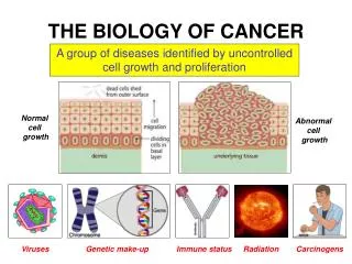
Cancer Biology. Tumour Markers Prof. Kenneth Bagshawe FRS. Tumour Markers Widest Definition. The concept of tumour markers can mean anything that helps in the diagnosis of cancer In a more restricted sense it refers to the biochemical detection of cancer. Imaging Techniques.

E N D
Cancer Biology Tumour Markers Prof. Kenneth Bagshawe FRS
Tumour MarkersWidest Definition • The concept of tumour markers can mean anything that helps in the diagnosis of cancer • In a more restricted sense it refers to the biochemical detection of cancer
Imaging Techniques Detected by: x-ray +/- contrast media Ultra sound Computerised Tomography (CT) Magnetic resonance imaging (MRI) Gamma Camera and radioisotopes (Nuclear Medicine)
Detection of Tumour Markers • Those detected on cancer tissue i) Biochemical methods Oestrogen receptors in breast cancer ii) Genetic methods Mutated genes BRCA1 and BRCA2 in breast cancer iii) Antibody methods Carcinoembryonic antigen (CEA)
Secreted by cancers into the blood Detection by: • Biochemical techniques steroids in adrenocortical cancer • Immunoassay proteins, glycoproteins
Ancient History • 1848 Bence Jones Multiple myeloma • 1929 Ascheim-Zondek hCG-trophoblast • 1932 Harvey Cushing Pituitary Basophils
The First Tumour Marker • It is known as Bence –Jones proteinuria, a marker for multiple myeloma a malignancy of the plasma cell. Often referred to as a paraproteinuria • It is a monoclonal gammopathy with any class of immunoglobulin
Adrenocortical Phaeochromocytoma Insulinoma Gastrinoma Carcinoid Medullary Ca Thyroid Cortisol adrenaline & nor adrenaline insulin, C peptide gastrin 5-HIAA Calcitonin Cancers of Endocrine Organs
Ectopic product syndromes • Cancers sometimes produce substances inappropriate to the cell type of origin • Bronchial carcinomas may produce ACTH, parathormone and human Chorionic Gonadotrophin (hCG)
Protein markers of common cancers detected by immunossay in blood • Human Chorionic gonadotrophin (hCG) Pregnancy Trophoblastic disease - Choriocarcinoma Germ cell cancers of testis and ovary • Alfa- feto protein (AFP) Germ cell cancers of testis and ovary Hepatoma
Protein markers continued • Carcinoembryonic antigen (CEA) Colon, stomach etc • Prostate Specific antigen (PSA) CA125- Ovary CA153- Breast
Measurement of Tumour Markers • 1930-1963 Bioassays on Urine • 1958 Immunoassays- serum • 1960 Radioimmunoassay- serum or urine • 1968 Immunometric assay serum or urine- Commercial Kits • 1975 Dip Stick Tests
What Is An Assay ? Unknown
Antibody Production Immunogen (Antigen) Rabbit Sheep Antiserum Mouse Horse etc Antisera contain many different antibodies directed at one or more sites on immunogen (polyclonal) Burnett (1957)- Clonal selection Theory One Cell - identical antibodies
Antibody Production • Kohler & Milstein (1975) Immortalise an antibody producing cell by fusion with a malignant cell • Hybridoma Hybridomas proliferate Identical (monoclonal) antibodies
RISING levels of these serum markers indicate disease progression • FALLING levels and slowing of rate of increase may indicate response to therapy • BUT interpretation has to take account of the HALF -LIFE (t1/2)
Half Life Of Common Tumour Markers Depends On Molecular Weight • MW TM T1/2(D) ROUTE • <70 hCG, PSA 2.3 renal • 70 aFP 6 mixed • > 70 all others 10 hepatic
Median Doubling Timesreflect different growth rates of cancers • CANCER TM T2(D) • Choriocarcinoma hCG 3 • Germ cell aFP 6 • Breast CA153, CEA 30 • Ovary CA125 30 • Pancreas CA199 30 • Colon CEA, CA199 30 • Prostate PSA 128
Human Chorionic Gonadotrophin (hCG) Normal Pregnancy: Mostly Intact molecule (α and β subunits) Mole, Choriocarcinoma: Fragmented hCG molecules
Therapeutic Application Radiolabelled Antibodies (RIT) Drug-Antibody Conjugates
Problems • Only a fraction of the i.v injected antibody gets into tumour: Most of the antibody stays in blood until excreted or metabolised • Heterogeneity of cancer cell: not all cancer cells express the marker
Potential Solutions • Antibody directed enzyme prodrug therapy (ADEPT)