Download
1 / 37
370 likes | 494 Vues
Crystallogaphy -- lecture 25. Protein guided tours: the meaning of “ Life ”. All life is based on reduced carbon. anoxic. oxidizing. 4*10 9 y. 3*10 9. 2*10 9. 1*10 9. present. origin of life. Chloroplasts. origin of oxygenic phototrophs . oxygenated environment.

E N D
Crystallogaphy -- lecture 25 Protein guided tours: the meaning of “Life”
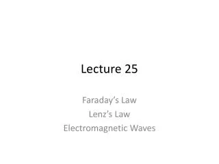
All life is based on reduced carbon anoxic oxidizing 4*109 y 3*109 2*109 1*109 present origin of life Chloroplasts origin of oxygenic phototrophs oxygenated environment Thanks to Doug Whittet, RPI Physics
O2 in the atmosphere provides a strong natural selection for CO2 fixation A narrowly avoided catastrophe for life on earth: • Cyanobacteria evolved a means to use light for energy, with O2 as the by-product. PHOTOSYSTEM I • O2 oxidizes everything in sight. • No reduced carbon left. • Everything would have died if not for Rubisco/Nitrogenase!! chloroplasts are descendents of the early cyanobacteria.
Classes of membrane proteins • Single transmembrane helix • several transmembrane helices • beta-barrel or channel • Anchored by one (not-transmembrane) helix or a covalently attached fatty acid
Photosystem I: Guided tour Download and display 1JB0.pdb (one jay bee zero) restrict !protein and !hohcolor cpkDisplay -> ball and stickselect magnesiumlabel %rset fontsize 12set fontstroke 2color labels yellow Find the pseudo 2-fold axis How many Mg are there? What are the residue numbers of the “special pair” of chlorophylls?
Photosystem I : Guided tour (select the special pair using select XXX or YYY)spacefillselect hetero and !hohlabels offcolor temperature How are the B-factorsdistributed? Was NCS 2-fold symmetry enforced during refinement? Which side is more ordered? Chain A or chain B? Guess what: 2-fold symmetry was not enforced during evolution!
Photosystem I : Guided tour Find the name of the lipid that does not havea phosphate group. Unix shortcut: use grepgrep ^”HETNAM” 1JB0.pdb Characterize the environmentof the lipid. Could it have a role in the light harvest process? select [LMG]restrict selectedcenter selectedselect within (11., [LMG]) and proteinDisplay -> ball_and_stickcolor cpkselect within (11., [LMG]) and ligand
Photosystem I : Guided tour select within (11., [LMG]) and ligandspacefill 1.5color green select within (11., [LMG]) and *.MGspacefill 1.5color white select within (11., [LMG]) and [PQN]color red select within (11., [LMG]) and solventspacefill 1.0color cyan What is PQN?How close is it to the nearestmagnesium
Photosystem I : Guided tour restrict ligandwireframecolor cpkDisplay-> ball and stickselect [CL1] or [CL2]wireframe color greenselect [PQN]color magentaspacefill 1.0select [BCR]color orangespacefill 1.5select *.MGspacefill 1.0color white Light harvesting complex Trace the path of the electronsfrom the special pair to the twoquinones. Are are of the pigmentsconnected to the special pair?
Photosystem I : Guided tour restrict [PQN]spacefill color cpkselect within (11.,[PQN]) and proteinwireframe 0.5color cpkselect within (11.,[PQN]) and ligand and not [PQN]color greenwireframe 0.5select within (11.,[PQN]) and solvent spacefill 0.6color cyan Environment of the quinones Which quinone is more loosely-bound How does the electron getfrom one quinone to theother? What protein sidechainforms a bridge?
Photosystem I : Guided tour Draw a TOPS diagram of chain D residues 24-95 only Look in scop.berkeley.edu for the fold under alpha+betaWhat fold is this?
Rubisco RUBISCO = Ribulose Bisphophate Carboxylase/Oxygenase Rubisco Animals ---> sugars, etc.
Rubisco fixes CO2 5 + 1 = 3 + 3 One of the substrates is small and feature-less. Gets confused with O2.
Competing oxygenase activity normal reaction Unwanted competing reaction
RUBISCO-FAQ • Rubisco is notoriously inefficient. Why?Oxygenase activity is an unwanted side-effect, but unavoidable since O2 is of similar shape and is a better electron sink than CO2. Also, CO2 has a lower partial pressure in the atmosphere. • Carbon fixation evolved exactly once. Right?Right. Otherwise we would see multiple proteins having this function. Rubisco must have been an enormous advantage over its contemporary competition. • How does rubisco overcome the inefficiency problem? Rubisco forms large complexes in order to have a larger concentration in the cell. • Why does rubisco require chaperonins to fold?The ultra-high concentrations of rubisco in the chloroplasts mean that misfolded rubisco quickly aggregates. Chaparones prevent aggregation.
Guided tour: 8RUC xtal symmetry Download 8RUC.pdb from www.rcsb.org rasmol 8RUC.pdb color --> chain display--> cartoons set unitcell on 8RUC space group is C2221 What crystal class is this?
Guided tour: edit the pdb file edit 8RUC.pdb (vi or jot) Note resolution of the data. Number of reflections. Number of atoms. Number of solvent atoms. Find “HETNAM” records. These are the “hetero” groups. Find the CAP and KCX “ATOM” records. Check the B-factors. How well ordered are they?
Guided tour: the A chain Rasmol commands: restrict :A center :A display-->cartoons stereo -7 Adjust the window to eye separation. Relax eyes to see stereo. Trace the chain from N to C. How many domains are there? What “fold” is each domain? Find it in SCOP and/or CATH.
Guided tour: the active site Rasmol commands: restrict within (10., 201:A) center within (10., 201:A) wireframe 50 color-->CPK select 201:A | CAP:A wireframe 80 select hetero & within (10., 201:A) & MG spacefill 1.0 select hetero & within (10., 201:A) & HOH spacefill 0.5 options-->slab mode
Guided tour: the active site (cont’d) Which residues are coordinating the magnesium? Where is the CO2 that was added to the ribulose? Rasmol commands: select :A & not within (10., 201:A) options-->slabmode (off) display-->backbone color-->group (this colors from N to C) Where is the active site relative to the domain? select not :A & protein display-->backbone color --> chain Where is the active site relative to the oligomeric contacts?
Guided tour: protein-protein contacts select not :A & within (10., :A) display-->sticks color red What chains are in contact with the A chain? What residues are involved? What command would select only waters that arewithin 5A of both chain A and chain L?
Nitrogenase 1N2C anoxic oxidizing 4*109 y 3*109 2*109 1*109 present Like CH2, NH3 was plentiful before oxygenic phototrophs. Since then, most N in the atmosphere is in the form of N2. Nitrogenase converts N2 to NH3
Nitrogenase is a hetero-tetramer. 2 Fe-proteins, 2 MoFe-proteins. With 3 Iron-containing clusters. Fe-protein MoFe-protein The Fe-protein cluster passes e- to the P-cluster, which passes them to the FeMo-cluster. e- accumulate at the FeMo-cluster, where the reduction of N2 takes place.
There are two Fe-containing clusters in the MoFe-protein FeMo cluster Reduced P-cluster Oxidized P-cluster dithionite A protein conformational change might favor oxidized over reduced, driving the reaction forward. from Rees & Howard (2000) Current Opinion in Chemical Biology, 4(5):p559-566.
Mechanism: simplified 2ADP dissociation of complex is rate-limiting 2ATP slow ATP ATP Fe-protein (red) ADP ADP Fe-protein (ox) +2Pi MoFe-protein (red) + fast MoFe-protein (ox) less than 6 e-? N2 + 6H+ H2 This reaction won’t happen until there are an accumulated 6e– 2H+ NH3
Mechanism: over-simplified Conformational changes couple hydrolysis of ATP with oxidation potential of Fe in Fe-protein 6e- 2NH3 N2 Fe2+distorted FeMo oxidized ATP MoFeprotein Fe-protein ADP FeMo reduced Fe2+undistorted
The high cost of nitrogen fixation The stoichiometry of nitrogenase is still not completely known. N2 + (6+2n)H++ (6+2n)e– + p(6+2n)ATP--> 2NH3 + nH2 + p(6+2n)ADP + p(6+2n)Pi n=number of H2 molecules formed (1 or 2, unknown) p=number of ATP required per electron (probably 2) H2 2H+ oops. nitrogenase
Many small molecules bind to the FeMo-cluster O2 (molecular oxygen) inactivates CO (carbon monoxide) inhibits (competitive) HCCH (acetylene) substrate HCN (cyanide) substrate N3- (azide) substrate N2 binds with KM = 0.02 atm
Interesting mutants of nitrogenase His 195A --> Glu Blocks N-fixation but allows reduction of acetylene. Gly 69A --> Ser Blocks reduction of acetylene, but allows N-fixation.
Guided tour of Nitrogenase (1N2C) Download 1N2C.pdb from www.rcsb.org In RasMol answer the following questions using the scripts provided plus any additions of your own: load 1N2C.pdb Display-->backbonecolor-->chainselect 50 and alphalabel %cset fontsize 20set fontstroke 4color labels yellow line up the molecule along the non-crystallographic 2-fold. There are 8 chains in the asu. Which chains are related to which by non-crystallographic symmetry?
Guided tour of Nitrogenase (1N2C) labels off select 68-69:Awireframe 50restrict selectedcenter selectedcolor cpk Line up atoms 69:A n and 69:A ca to measure the phi angle. R-handed is positive. Does Gly69A have a positive phi-angle? Mutating G69 blocks reduction of acetylene, but allows N2-fixation. Would mutating Gly69A to a Serine possibly change its conformation?
Guided tour of Nitrogenase Draw a TOPS diagram of chain E. restrict :Ecenter selectedDisplay-->cartooncolor-->structureLine the structure up with the beta sheet perpendicular to the screen. Ignore short helices (they are not really helices). Draw strands as up or down arrows and the helices as circles. Then draw connecting lines, to the middle if the connection is toward you, to the edge if the connection is away from you. Find the N-term. Number the strands from N to C. Find the fold class from SCOP (scop.berkeley.edu). (goto “top of the heirarchy” then class 3, “alpha and beta proteins”)
Finding the fold name in SCOP Chain E is “3-layer”. When you have numbered the strands, look in SCOP for the “Fold” with the observed strand order (for example: 43125, etc). (cute trick: use the browser’s “search in page” function). The strand order can be read from right-to-left or left-to-right. If a terminal strand is at the edge of the beta sheet it might be missing. Also, extra strands might be added at the C-term or N-term is it occurs at the sheet edge. Write the fold name here ______________________
Guided tour of nitrogenase Characterize the environment of the ADP and metal clusters. select within (6., hetero) and (:A | :B | :E | :F)restrict selectedDisplay -->ball and stickcolor whiteselect selected and heterospacefillcolor cpkselect within (6., hetero) and (:A | :B | :E | :F)select selected and acidiccolor red select within (6., hetero) and (:A | :B | :E | :F)select selected and basiccolor blue continued...
Guided tour of nitrogenase Characterize the environment of the ADP and metal clusters. select within (6., hetero) and (:A | :B | :E | :F)select selected and polar and not basic and not acidiccolor green How would you characterize the binding sites? (check one for each het group) mostly mostly mostly mixed non- basic acidic polar charges polarADPFS4CLFCFMCA