Download
1 / 25
290 likes | 482 Vues
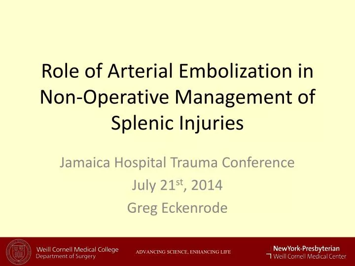
Role of Arterial Embolization in Non-Operative Management of Splenic Injuries. Jamaica Hospital Trauma Conference July 21 st , 2014 Greg Eckenrode. ADVANCING SCIENCE, ENHANCING LIFE. Management of Traumatic Spleen Injuries. Historically, nearly all splenic injuries were managed operatively

E N D
Role of Arterial Embolization in Non-Operative Management of Splenic Injuries Jamaica Hospital Trauma Conference July 21st, 2014 Greg Eckenrode ADVANCING SCIENCE, ENHANCING LIFE
Management of Traumatic Spleen Injuries • Historically, nearly all splenic injuries were managed operatively • Non-operative management developed in the pediatic population in the late 1960s • Increasing prevalence in adult population since the 1980s • Currently 50-70% of splenic injuries Stein DM, Scalea TM J Intensive Care Med. 2006;21(5):296.
Operative Management • Hemodynamically unstable patients with evidence of abdominal bleeding • Positive FAST or DPA/DPL • Patients requiring abdominal exploration for other injuries • Intraperitoneal free air • Signs of peritonitis
Operative Management • Hemodynamically stable patients • CT findings of contrast extravasation or vascular blush • High grade injuries (generally IV-V) • Age > 55 • Unable to safely observe patient
Conventional Non-Operative Management • Admit to monitored care setting • Bed rest, NPO • Serial Hgb/Hct every 6 hours for 24 hours • Frequent vital signs, serial abdominal exams
Splenic Angiography and Embolization • First applied to traumatic splenic injuries in 1995 • Multiple techniques • Distal selective • Proximal • Both • Intended to improve success of non-operative management
Clinical Questions • Does splenic artery angiography and embolization improve non-operative management outcomes? • Which patients should undergo angiography and embolization?
Western Trauma Association • 4 L1 trauma centers in the United States • 155 patients who underwent angiography and embolization for pseudoaneurysm, active bleeding on CT, significant hemopertoneum, and high grade injuries • Compared against the results of the Eastern Trauma Association study, which used conventional observation
Comparison Results Haan, et al; J Trauma. 2004 Mar;56(3):542-7.
Ullevaal University Hospital: 2006 • In 2002, implemented policy that all patients with splenic injury Grades III-V or ongoing bleeding underwent arterial embolization • Compared to all splenic injuries from 2000-2002, when arterial embolization was no performed at the hospital Gaardner, C, et al; J Trauma. 2006 Jul;61(1):192-8
Multicenter Variation: 2010 • Compared 4 L1 trauma centers with variation in rates of splenic artery embolization in non-operative management • Rates ranged from 19% to 1% • Compared rates of splenic salvage and non-operative failure Bannerjee, et al; J Trauma Acute Care Surg. 2013 Jul;75(1):69-74
Population Comparison Bannerjee, et al; J Trauma Acute Care Surg. 2013 Jul;75(1):69-74
Management Comparison Bannerjee, et al; J Trauma Acute Care Surg. 2013 Jul;75(1):69-74
Splenic Salvage Rate Bannerjee, et al; J Trauma Acute Care Surg. 2013 Jul;75(1):69-74
Wake Forest - 2014 • Single site L1 Trauma Center • Prior to 2010, angiography and embolization performed for CT contrast blush • Starting in 2010, prospectively performed angiography and embolization on all Grade III-IV splenic injuries • Compared non-operative failure rates against recent historical controls from 2007-2009 period Miller, et al; J Am Coll Surg. 2014 Apr;218(4):644-8
Study Group Comparison Miller, et al; J Am Coll Surg. 2014 Apr;218(4):644-8
Study Results • 2010-2012: Non-operative failure rate of 5% • Failure rate of 25% in 16 protocol deviations (p=0.02) • 2007-2009: Non-operative failure rate of 15% (p=0.04)
Conclusions • In historical comparisons, patients with splenic injuries who are candidates for non-operative management have better outcomes when SAE is utilized • Centers which perform a higher rate of SAE have higher rates of spelic salvage and lower rates of non-operative management failure
Conclusions • Centers which implement a standard protocol mandating SAE for non-operative splenic injuries experience decreased rates of of non-operative failure and increased rates of splenic salvage
Future Directions • No prospective, head-to-head randomized clinical trial of SAE in non-operative splenic injuries • Limited data with respect to cost effectiveness
Splenic Injury Grading • Grade I: • Hematoma: Subcapsular, < 10% of surface area • Laceration: Capsular tear < 1 cm depth • Grade II • Hematoma: Subcapsular, 10 - 50 % of surface area • Laceration: Capsular tear 1 - 3 cm depth not involving trebecular vessel
Splenic Injury Grading • Grade III • Hematoma • Subcapsular, > 50% of surface area • Subcapsular, expanding • Ruptured subcapsular or parenchymal • Intraparenchymal > 5cm • Laceration • > 3cm depth • Involving trabecular vessel
Splenic Injury Grading • Grade IV • Laceration: segmental or hilar vessels with > 25% devascularization • Grade V • Hematoma: shattered spleen • Laceration: total devascularization