Download
1 / 39
631 likes | 1.61k Vues
RH-ISOIMMUNIZATION AND & ABO incompatibility. Prepared by N. Bahniy. Rh- Iso imunization Definition. known as: Rhesus incompatibility , Rhesus disease RhD Hemolytic Disease of the Newborn.

E N D
RH-ISOIMMUNIZATION AND & ABO incompatibility Prepared by N. Bahniy
Rh- Iso imunizationDefinition known as: Rhesus incompatibility, Rhesus diseaseRhD Hemolytic Disease of the Newborn. -When Rh– mother gets pregnant to Rh+ fetus —she may be sensitized to Rh antigen and develop antibodies. These will cross the placenta and cause hemolysis of fetal red blood cells. - The risk of sensitization after ABO incompatible pregnancy is only 2%
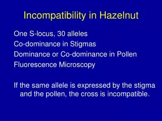
Pathophysiology The rhesus system which comprises number of antigens C, D, E, c, e. A person who lacks D antigen is called Rh negative. 15% of Caucasians, 5% African Americans and 2 % of Asians are Rh negative. Rh isoimmunisation is due to D antigen in more than 90% of cases. Occasionally hemolytic disease of the newborn is a result of maternal immunization to Irregular RBC antigens other than Rh group like anti- Kell and anti- Duffy
Pathophysiology Initial response is forming IgM antibodies for short period followed by production of IgG which crosses placenta IgG antibodies adhere to the antigen site on the surface of erythrocytes causing hemolysis. The excessive removal of circulatory RBCs leads to severe anemia and hypoxia. Erythropoiesis results inhepatosplenomegaly. Tissue hypoxia and hypoproteinemia results in cardiac and circulatory failure, with generalized odema and hydrops
Mother 1. Cleared by Macrophage Primary Response 2. Plasma stem cells • 6 wks to 6 M. • IGM. IGM antibodies Placental Fetal Anaemia
Mother Macroph. antigen Presenting cell T- helper cell Secondary Response • Small amount • Rapid • IgG B cell IgG Anti - D Placental Fetal Anaemia
Mother Macroph. Antigen Presenting Cell Group “O” Rh Negative T-Hellper Anti - A Anti - B B-cell Anti-D Placenta A Rh positive B Rh Positive Infant “O” Rh positive
7% in the first trimester. 16% in the second trimester 29% in the third trimester Risk of fetromaternal hemorrhage is increased in abruption placenta, threatened abortion, toxemia, after cesarean section, ectopic pregnancy, amniocentesis, intrauterine fetal transfusion. And it occur during normal delivery Fetomaternal hemorrhage as a reason of Rh –isoimmunization has been documented in:
Rh Antibodies Antibodies Coated Red Cells Destruction of Fetal Cells by Fetal RES Fetal Anemia Fetal Hypoxia and Stimulate of Erythropoitin Extra Medullary red Cells Synthesis Hepatomegally Hepatic Cell Failure Hypoproteinemia, Increased Intrahepatic Pressure, Portal hypertension Ascetic, Edema, hypoxia, Placental Thickness, Polyhydramnios, Pericardial effusion
Complications of Fetal-Neonatal anemia: • Fetal Hydrops And Stillbirth • Hepatosplenomegaly • Neonatal Jaundice • Compilations Of Neonatal Kernicterus (Lethargy, Hypertonicity, Hearing Loss, Cerebral Palsy And Learning Disability) • Neonatal Anemia
Kernicterus Concentration of bilirubin in the newborn blood exceeds in-term fetus – 307,8 – 342 mkmoll/L in pre-term fetus – 153-205 mkmoll/L,
Natural History • 50% of affected infants have no or mild anemia, requiring either phototherapy or no treatment. • 25% have some degree of hepatosplenomegaly and moderate anemia and progressive jundice culminating in kernicterus, neonatal death or severe handicap. • 25% are hydropic and usually die in utero or in the neonatal period ( half of these the hydrops develops before 34 weeks gestation
To predict which pregnancy is at risk To predict whether or not the fetus is severely affected. To correct anemia and reverse hydrops by intrauterine transfusion. To deliver the baby at the appropriate time, weighing the risks of prematurity against these of intrauterine transfusion. The aim of antenatal management
First ante-natal visit check blood group, antibody screening. If indirect coombs test is positive, the father’s Rh should be tested. Serial maternal Anti D titers should be done every 2- 4 weeks. If titer is less than 1/16 the fetus is not at risk. If titer is more than 1/16 then severity of condition should be evaluated. Recognition of pregnancy at risk
Prediction of the severity of fetal hemolysis History of previous affected pregnancies The levels of maternal hemolytic antibodies Amniocentesis Biophysical surveillance Fetal blood sampling
- There is an excellent correlation between the amount of bilirubin in amniotic fluid and fetal hematocrit. - the optical density deviation at 450 nm measures the amniotic fluid unconjugated bilirubin. Amniocentesis – at 16 weeks Ultrasound image of amniocentesis at 16 weeks of gestation
Amniocentesis • Normally Bilirubin In Amniotic Fluid Decreases With Advanced Gestation. • It Derives From Fetal Pulmonary And Tracheal Effluents. • Its Level Rises in Correlation With Fetal Hemolysis. Determination Of Amniotic Fluid Bilirubin: By The Analysis Of The Change In Optical Density Of Amniotic Fluid At 450 nm On The Spectral Absorption Curve (delta OD450) Procedures Are Undertaken At 10-15 Days Intervals Until Delivery Data Are Plotted On A Normative Curve Based Upon Gestational Age.
Ultrasound detection of Rh Sensitization - Placental size and thickness and hepatic size. - Fetal hydrops is easy to diagnose when finding one or more of the following: Ascites, pleural effusion, pericardial effusion, or skin edema. - Doppler assessment of peak velocity of fetal middle cerebral artery proved to valuable in predicting fetal anemia
Rh- Iso imunization Body wall edema hydropic fetus
Rh- Iso imunization Fetal Ascites
Biophysical surveillanceMiddle cerebral artery peak velocity
Biophysical surveillanceMiddle Cerebral Artery peak systolic velocity A = moderate-severe anaemia B = mild anaemia C = no anaemia 80 70 60 50 40 30 20 A 1.5 MOM B 1.29MOM MCA peak velocity cm/sec C Median 20 25 30 35 Gestational Age (wks) from Mari et al, NEJM 2000; 342:9-14
Cordocentesis - Diagram of cordocentesis procedure
Percutaneous Fetal Blood Sampling - allows measurement of fetal Hb, Hct, pH, reticulocytes
Is the gold standard for detection of fetal anemia. Reserved for cases with: - With an increased MCA-PSV
Monthly Maternal Indirect Coombs Titre Complicated History and / or Exceeds Critical Titre Below Critical Titre Paternal Rh Testing Rh Positive Rh-negative Amniocentesis for RhD antigen status Routine Care Fetus RhD positive Fetus RH D Negative Weekly MCA-PSV Serial Amniocentesis > 1.50 MOM < 1.50 MOM Cordocentesis or Deliver Suggested management of the RhD-sensitized pregnancy
Serial Amniocentesis Lily zone I Lower Zone II Zone III Hydramnios & Hydrops Upper Zone II Repeat Amniocentesis every 2-4 weeks < 35 to 36 weeks And Fetal lung immaturity > 35 to 36 weeks Lung maturity present Delivery at or near term Intrauterine Transfusion Repeat Amniocentesis in 7 days or FBS Delivery Hct < 25% Hct > 25% Intrauterine Transfusion Repeat Sampling 7 to 14 days Suggested management after amniocentesis for ΔOD 450
Pregnant women undergo cesarean section in isoimunization: • Severe form of hemolytic infant disease in the term 34-35 weeks after previous antenatal prevention of fetal hyaline membranes syndrome; • Hydrops fetalis in any gestation term because of interm pregnancy would provoke antenatal fetal death.
In the second stage of labor pudendal block and episiotomy are indicated (they decreasing fetal trauma). In the all others cases pregnant women with the diagnosis of Rh- disease undergo delivery in the term of 37-38 weeks of gestation. Induction of labor is performen by prostaglandin (in the case of “unripe” uterine cervix) or by intravenous oxytocin infusion administration (in the case of “ripe” uterine cervix). Vaginal delivery in Rh-isoimmunization
Rh- Iso imunizationPrevention - Screening of all pregnant mothers to Rh D antigen and antibody screening for Rh D negative mothers. -Prophylactic anti D immunoglobulin to all Rh – mothers after delivery if the fetus is Rh+ or( at 28, 36 weeks of pregnancy) and after abortion, amniocentesis, abruption.
Rh- Iso imunizationPrevention The standard dose of anti D is 0.3 mg —will eradicate 15 ml of fetal red blood cells (routine for all Rh –ve pregnancies) within 3 days of delivery. -If more feto-maternal bleeding is suspected as in abruption or ante partum hemorrhage-Do Kleihauer –Betke test to estimate the amount of fetal red cells in maternal circulation and re-calculate the dose of the anti-D.
Management of sensitized newborn Mild anemia (Hb <14gm/dl, cord bilirubin>4 mg/dl)---Phototherapy -Moderate to severe----Exchange transfusion. -Mild Hydrops improves in 88% of cases -Severe hydrops—Mortality is 39%