Download
1 / 68
680 likes | 808 Vues
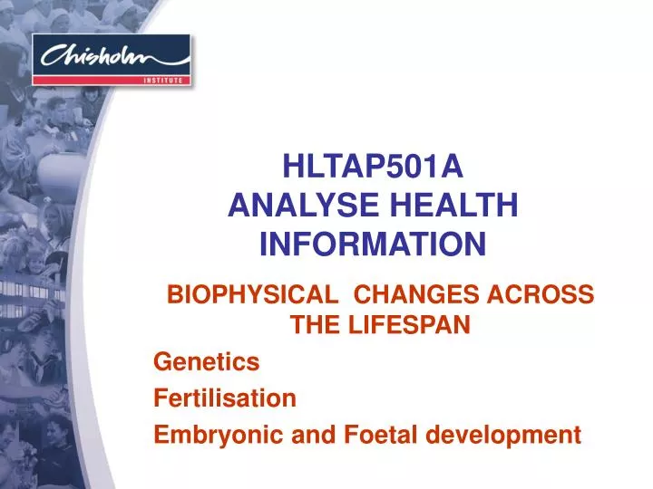
HLTAP501A ANALYSE HEALTH INFORMATION. BIOPHYSICAL CHANGES ACROSS THE LIFESPAN Genetics Fertilisation Embryonic and Foetal development. GENETICS. Genetics is a branch of biology that is concerned with: - How the traits of anatomy, physiology and behaviour is passed on to descendants

E N D
HLTAP501AANALYSE HEALTH INFORMATION BIOPHYSICAL CHANGES ACROSS THE LIFESPAN Genetics Fertilisation Embryonic and Foetal development
GENETICS • Genetics is a branch of biology that • is concerned with: - • How the traits of anatomy, physiology and behaviour is passed on to descendants • How the individual expresses those traits during development and their life.
Individuals inherit characteristics from their genetic parents : - • Hair & eye colour • Stature • Shape • Familiar traits • Susceptibility to genetic disease.
FERTILIZATION • Single cell, the fertilized ovum contains all inherited information which determines the individual’s characteristics and any possible inborn error of metabolism. • Genetic material is mainly in the cell nucleus and is passed on during cell division to ALL new cells.
http://www.yorku.ca/kdenning/++2140%202006-7/2140-17oct2006.htm (accessed 21/4/08)
CELL NUCLEUS • The cell nucleus contains chromosomes, which consist of long coils of deoxyribonucleic acid (DNA) in close contact with protein molecules. • The genes are organised in a linear array along the chromosomes, they are responsible for virtually all inborn and inherited characteristics
CHROMOSOMES • Humans have 46 chromosomes : - • 23 matching (homologous) pairs. • 22 pairs (44 chromosomes) are similar in both males & females and are called autosomes. • The pairs are number by length chromosome 1 being the longest & 22 the shortest.
http://www.yorku.ca/kdenning/++2140%202006-7/2140-17oct2006.htm (accessed 21/4/08)
SEX CHROMOSOMES • The remaining pair are the sex chromosomes. • In females they consist of two X chromosomes. • In males there is one X chromosome & one much smaller chromosome called the Y chromosome.
CELL DIVISION - Mitosis • Somatic cells (e.g., cells of the body) divide by a method of mitosis • This is a process by which the cell repairs & replace themselves. • This process retains all of the 46 chromosomes & creates an exact replica of itself
http://www.accessexcellence.org/RC/VL/GG/mitosis.php (accessed 21/4/08)
CELL DIVISION - Meiosis Germ cells (reproductive cells or gametes) divide by meiosis, this is a process where the number of chromosomes is halved (23). Fertilization of the ovum by the sperm restores the cell to it’s full complement of 46 chromosomes.
http://www.yorku.ca/kdenning/++2140%202006-7/2140-17oct2006.htm (accessed 21/4/08)
Meiosis – first division • Meiosis involves two nuclear divisions with only one doubling of genetic material. • At the first meiotic division, the two members of each pair of homologous chromosomes split into two daughter cells. This is called reduction division because the chromosome number is reduced to 23 (haploid)
The normal cell is diploid as it contains two of each chromosome. • Rarely a child is born with triploid, having three of each chromosome (69 in all)
Meiosis – second division • The second division is similar to the first except there are only 23 chromosomes to start with. • In each cell the 23 chromosomes split lengthways. The result is four daughter cells, each with 23 chromosomes. • The full diploid complement is restored at fertilization when the ovum and spermatozoa fuse
Meiosis differs from mitosis in that crossing-over can occur between chromosomes – this does not occur in mitosis. • Cross-over results in a shuffling of the genes on a pair of homologous chromosomes so that the chromosome passed on to the child from the parent ‘s mother nor the one from his/her father but a reassortment of these. • These recombination events are called chiasmata
CHROMOSOMAL ANOMALIES • Too many or too few chromosomes, or an alteration in their structure may result in spontaneous abortion. • Structural changes may result in a loss of a piece of chromosome (deletion). • Translocation is when a piece of chromosome breaks off and is joined to another. • Reciprocal translocation occurs when two chromosomes lose pieces and are joined to the wrong chromosome • The presence of just one chromosome is called monosomy, the presence of three instead of the usual two is called trisomy
COMMON ANOMALIES • Most common is Down’s Syndrome (47 instead of 46 chromosomes) often termed as trisomy 21. • The others are of the sex chromosomes and carry one X chromosome without a Y chromosome (monosomy X, Turner’s Syndrome) few foetuses survive • Other anomalies include XXX (girls) and XYY (boys)
INHERITANCE • Genetic disease is inherited in three different ways:- • Autosomal dominant inheritance • Autosomal recessive inheritance • 3. Sex linked
Inherited conditions are often described as being “dominant" or “recessive” (abbreviations for autosomal dominant and autosomal recessive) • The gene that dominates is called the dominant gene. • The trait is carried on one pair of the 22 homologous chromosomes. • The person who carries the defective gene has the disease/trait – this person is not a carrier
DOMINANTLY INHERITED CONDITIONS • Only one parent has to have a defective gene to produce children with dominantly inherited conditions. These include • Marfan’s Syndrome • Huntington’s Disease • Neurofibromatosis (Elephant man) • Achondroplasia (dwarfism)
http://craniofacialcenter.uiowa.edu/center/vws.php (accessed 21/4/08)
RECESSIVE CONDITIONS • The inhibited (hidden) gene is called a recessive gene, the recessive trait is carried on one of a pair of chromosomes. • If only one gene is defective the person is a carrier and does not exhibit any symptoms of the disease.
RECESSIVE CONDITIONS • Both parents must carry the defective gene to produce children with recessively inherited conditions. Most enzyme deficiencies are recessive conditions however if only one parent carries the condition the body’s metabolism can function adequately with reduced levels of most enzymes.
RECESSIVE CONDITIONS • Phenylketonuria (PKU) • Galactosaemia (error in galactose metabolism) • Cystic fibrosis • Sickle cell disease • Polycystic kidney disease
SEX-LINKED CONDITIONS • Carried on the X chromosome. These conditions are more complex because the gene dose varies between males (X) & females (XX). • In females only one of the two X chromosomes is active, which X is random through out the body. • In males are affected even by recessive disease which they carry on their X chromosome (women are not)
SEX-LINKED CONDITIONS • Sex linked recessive diseases are carried by the female and are passed on to their sons e.g., • Red-green colour blindness • Haemophilia • Duchenne muscular dystrophy
Sex-Linked Inheritance http://www.goldinfo.org/education.aspx (accessed 21/4/08)
The inheritance diagram shows possible outcomes for thechildren of:a haemophiliac father and normal mother, and a normal father and carrier mother. http://www.bbc.co.uk/schools/gcsebitesize/biology/variationandinheritance/geneticdiseasesrev3.shtml (accessed 21/4/2008)
SEX-LINKED CONDITIONS • A female who carries such a disease on one of her two X chromosomes will transmit it (on average) to 1 in 2 of her children – half of her sons will be affected & half of her daughters will be carriers. • A male will pass on his disease gene whenever he hands over his X chromosome. All his daughters will be carriers. His sons will not be affected as they receive his Y chromosome
FERTILISATION • Fusion of spermatozoon and ovum • usually occurs in fallopian tube • Once oocyte enters fallopian tube it has a lifespan of 6-24 hours • The spermatozoa can survive in a viable state for 30-80 hours
Fertilisation continued • During passage through genital tract, spermatozoa undergo final series of changes before they are ready for fertilisation. • Capacitation- the cell membrane undergoes a number of modifications including the removal of certain molecules added during ejaculation. • After capacitation spermatozoa exhibit hyperactive motility.
Fertilisation continued • After capacitation, the plasma membrane of the sperm & outer membrane of the acrosome fuse, the membrane breaks down, releasing enzymes that allow the sperm to enter the corona radiata. • Sperm digest their way through the zona pellucida via enzymes associated with the inner acrosomal membrane • Only sperm that have undergone arosome reaction are capable of fusing with the oocyte • Usually the first sperm to make contact with the oocyte proceeds to fertilisation. • In the process of fusion, the plasma membrane of the sperm head is phagocytosed by the oocyte
Day 1:fertilization: all human chromosomes are present; unique human life begins.Illustration by R.K. O'Bannon (www.nrlc.org/abortion/facts/fetaldevelopment.html [accessed 18/4/08])
ZYGOTE • The newly formed cell containing maternal & paternal chromosomes is called a zygote. • Within 2-3 hrs of fertilisation the zygote proceeds with the final phase of meiosis (that was halted immediately following ovulation) • In this phase the female chromosomes divide by mitosis, yielding one haploid set of female chromosomes within the main body of the cytoplasm (remaining set is discarded) • At the same time, the sperm contacts the cytoplasm content of the cell membrane & combines with that of the female and second meiotic division takes place. • Between 4-7 hrs following cell fusion, the 2 sets of haploid combine. • The pronuclei form and migrate toward • The first mitotic division transforms the zygote into a 2 cell conceptus. • Following the 2 cell stage, the pre-embryonic genome is activated
Approximately every 12 hours within the uterine tube a further series of undifferentiated cellular divisions occur until 8-16 cells have been formed. • At this point the conceptus reaches the uterine cavity where a new process of differentiated process of cellular division occurs. • At this stage the conceptus is called a morula (as it looks like a mulberry)
Blastocyst • Over the next 24 hrs the morula begins to accumulate fluid with in the centre, forming it into a thin layer of outside cells. • The blastocyst is made up of 34-64 cells. • At this stage the conceptus utilises metabolic substances from endometrial fluid for a further 24 hrs before the outer cells attaches itself to the uterine epithelium. • The outer rim of cells synthesis a glycoprotein hormone , chorionic gonadotrophin (hCG) which is similar to lutienising hormone (LH). Because of the similarity hCG acts on LH in thecal and granulosa cells of the corpus luteum • This causes continuous secretion of progesterone and oestrogen which maintains the endometrium in an optimum state for implantation.
http://www.biocrawler.com/encyclopedia/Mammalian_embryogenesis (accessed 21/4/08)
Trophoblast cells • Trophoblast cells make contact with the endometrium. These cells will differentiate in to 2 layers that form the beginning of the chorion & foetal portion of the placenta. • The chorionforms the wall of the chorionic sac • By day 9 the embryo is completely implanted into the uterine endometrium
www.jillstanek.com/archives/fertilization.jpg (accessed 18/4/08)
PLACENTAL FUNCTION • The placenta is an ORGAN that caters for all the requirements of the foetus in utero. • It selects these requirementsfrom maternal blood. • Many are used immediately, others are stored and or changed by the placenta to make them suitable for foetal use.
The placenta has five (5) main functions • Respiratory • Nutritive • Excretory • Protective • Endocrine (secretes every hormone)
UTERINE BLOOD FLOW • In non pregnant women uterine blood flow is approx. 40ml/min which rises by 10ml/min during the first trimester. • Much larger increases occur during the second & third trimesters • By the end of pregnancy it rises to over 800ml/min.