Download
1 / 40
450 likes | 815 Vues
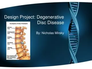
Degenerative disease. Aging and degeneration of intervertebral disc * this is a biochemical process, but it can be observed on MR * In children the nucleus pulposus has high signal on T2w and anulus fibrosus has a low signal * In adults the nucleus pulposus has high signal on

E N D
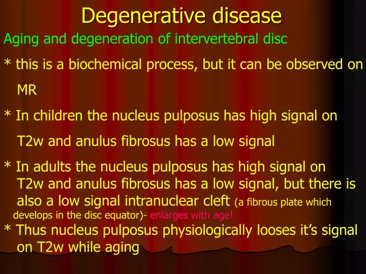
Degenerative disease Aging and degeneration of intervertebral disc * this is a biochemical process, but it can be observed on MR * In children the nucleus pulposus has high signal on T2w and anulus fibrosus has a low signal * In adults the nucleus pulposus has high signal on T2w and anulus fibrosus has a low signal, but there is also a low signal intranuclear cleft (a fibrous plate which develops in the disc equator)- enlarges with age! * Thus nucleus pulposus physiologically looses it’s signal on T2w while aging
Degenerative disease • Disc degeneration phase I • disintegration of mucopolisaccharides’ chains leads to • the higher water accumulation within the disc • intervertebral disc becomes thickened on plain films • and MR and have higher signal on T2w • on functional plain rtg. films the disc deformations • caused by movement are smaller • acute large disc herniations possible
Degenerative disease • Disc degeneration phase II • the lower content of large molecule proteoglycans and • mucopolysaccharides leads to dehydrtation of the • nucleus pulposus • loss of disc space height on plain films and MR and • signal on T2w • posterior and/or anterior bulging on CT and MR • means that anulus fibrosus is shifted outside the • vertebrae (posterior longitudinal ligament) line
Degenerative disease • Disc degeneration phase III • disintegration and framentation of nucleus pulposus • smaller disc height means narrowing of the intervertebral • space meansrelative anulus fibrosus fibers elongation • results inbulging disc • smaller disc height visible both plain films and MR • disc herniations visible on CT i MR • osteophytes and vertebral border plates hyperostosis on • plain films • instability and spondylolisthesis on plain films and MR
Degenerative disease • Disc degeneration additional signs • vacuum disk – intradiscal gas = CO2plain films, MR, CT • vertebral arc disruption = spondylolysis and • spondylolisthesis plain films, MR, CT • vertebral bone marrow changes – only MR!: • # type 1:inflammatory – like diskitis - low signal on T1w and high on T2w – damage of the vertebral border plates and highly vascular fibrous tissue, CE is visible • # type 2:high signal on T1w and high but not very on T2w – fatty marrow displacement – do not enhance after Gd
Disc degeneration phase II i III Spondylolysis and spondylolisthesis
phase II & III Fatty marrow displacement inflammatory lesions with diskitis
Degenerative disease What can cause a vertebral canal stenosis? • Bulging and herniation • Spondylosis and osteophytes • Thickening, calcification and ossification of posterior longitudinal and yellow ligaments • Arthrosis – degenerative changes in intervertebral joints, synovial cysts (mixed density and signal on CT and MR) • spondylolisthesis • Nondegenerative: Congenital – Achondroplasia, Morquio syndrome, Short pedicle syndrome Miscellaneous – Ossification and calcification of ligaments, epidural lipomatosis
Degenerative disease • Disc herniation on CT • hyperdense structure, rarely calcified, localized inside the spinal canal at the level or/and lower or above the level of the intervertebral space • the menimgeal sac, spine or sometimes nerve root compression can be visualized
Degenerative disease • Disc herniation in MR • features of anulus fibrosus disruption can be visible - anular tears – small hypreintense areas on T2w • - Later well seen discontinuity of the anulus • asymmetric protrusion of the disc material into the spinal canal (usually low signal on T1 & T2w) visible within the spinal canal and intervertebral foramina
Degenerative disease • Disc herniation in MR • direct visualisation of spine, meningeal sac and nerve roots compression • CE is visible around the acute herniations and postsurgical scars
Degenerativedisease • Disc herniation MR - postsurgical • MR enables to visualize problems directly after the surgery: epidural haematoma, residual disc fragments, inflammatory changes (but the differential diagnosis is not highly specific!!!) • postsurgical scars diagnosis • potsurgical scars differetial diagnosis with the reccurent disc herniation
Spine Tumors • Primary neoplasms are less common than brain • neoplasms • nerve sheath tumors are more common than neuroepithelial ones • Clasification of spine lesions by anatomic compartment • extramedullary • intramedullary
Spine Tumors Extramedullary tumors * extradural - osseous spine tumors - metastases - lipomas - Sometimes schwannomas and neurofibromas * intradural
Spine Tumorsextramedullary tumors • * intradural • - nerve sheath tumors (most common): • (schwannomas and neurofibromas) • meningiomas (second most common after nerve sheath) • cysts • lymphomas
Spine Tumors Extramedullary tumors * extradural most comon are metastases - affect and infiltrate every part and structure of the spinal canal - on plain films and CT osteolytic or osteosclerotic focal lesions
Spine Tumors Extramedullary, extradural metastases on MR perfect visualization of the lesion extent within all spinal canal structures - on T1 w the signal is lower than marrow and CE is present - on T2w the signal is higher than marrow but not as high as in fatty replacement foci
Meta – infilration of vertebral arcs, and processes, spinal canal mass
Spine Tumorsdifrential diagnosis of benign and pathologic vertebral fracture on MR Benign (osteoporotic, or trauma): • signal of the fractured vertebral bodies is similar to others – unbroken (high on T1w, low on T2w) • Relatively uniform • Multiple changes localized in the neighbouring vertebrae usually in the middle of physiological kyphosis or lordosis • Fat suppression techniques may be useful Malignant (metastases): • Multiple changes localized with wider distribution, not neighbouring • signal of the fractured vertebral bodies is different from unbroken ones (low on T1w, high high on T2w), • Osteosclerotic lesions have low signal both on T1& T2w • Signal is inhomogenous • CE present • All parts of the verterae can be infiltrated (body, arcs and proceses)
Spine TumorsExtramedullaryintradural • nerve sheath tumors: • Schwannomas • Neurofibromas • Ganglioneuromas • Neurofibrosarcomas common rare
Spine TumorsExtramedullaryintradural • Schwannoma, Neurofibroma • (can not be definitely differentiated using the diagnostic imaging) • - can be intra 70-75% or extradural up to 30% • - on CT only larger „dumbbell” type tumors are visible • they cause enlargement of intervertebral foramina and • come through them outside the spinal canal • hypo up to sligthly hyperdense on CT • calcifications and bleeding are rare
Spine TumorsExtramedullaryintradural • Schwannoma, Neurofibroma(can not be definitely differentiated using the diagnostic imaging) • onMR: T1w isointense to spinal cord (75%), 25% hypointense • on T2w hyperintense to spinal cord (95%), but hypo to CSF • Schwannomas in 40% inhomoenous (bleeding, calcifications, hyalination) • Neurofibromas „target sign” on T2w and T1CE • hyperintense rim(myxomatous tissue)and hypointense • center(fibrocartilagineous tissue) • contrast enhancement (CE) usually weaker than in meningiomas
Spine TumorsExtramedullaryintradural • meningiomas • middle aged women • location: thoracic spine • 90% intradural • oval shape • may be „dumbbell” type • on CT calcifications and hyperostosis can be visible • (en plaque meningioma)
Spine TumorsExtramedullaryintradural • meningiomas • - on MR complete imaging feature of the tumor, differential diagnosis is possible • on T1 & T2w sygnal is similar to spinal cord • on T1w strong homogenous enhancement, tumor well visible • tail sign may be visible • in calcified tumors, low signal both T1&T2w and no CE
meningioma T2W T1w CE
Spine Tumorsintramedullary • - gliomas: ependymoma and astrocytoma • there is no possibility to differentiate this 2 tumors • using diagnostic imaging • vascular lesions:haemangioblastoma, AVM • metastases • intramedullary cysts: hydrosyringomyelia, • inflammatory, malatic –post bleeding
Spine Tumorsintramedullary • ependymomas • - 60% of spine gliomas and 90% of the cauda equina and filum terminale gliomas • most common intramedullary tumor in adults • middle aged adults, female predominance • location: cauda equina and filum terminalemyxopapillary or cervical spineCellular (with lower incidence) • - onCTdifficult to visualize, unspecific spinal canal enlargement
Spine Tumorsintramedullary ependymomas MR • On T1w hypo or isointense to spinal cord • On T2w usually hyperintense • Moderately strong, inhomogenous CE • Cyst less common tha in astrocytomas • Bleeding more common than in astrocytomas
Spine Tumorsintramedullary • Astrocytomas • usually low grade fibrillary astrocytoma • most common spinal cord tumor in children, second common in the whole population • long multisement mass enlarging the spinal cord • enlarged spinal canal onplain films and CT • on MR iso to hypointenseg on T1w and hyperintense on • T2w, fusiform, multisegment enlargement of the cord • cysts common • always enhance