Download
1 / 16
170 likes | 346 Vues
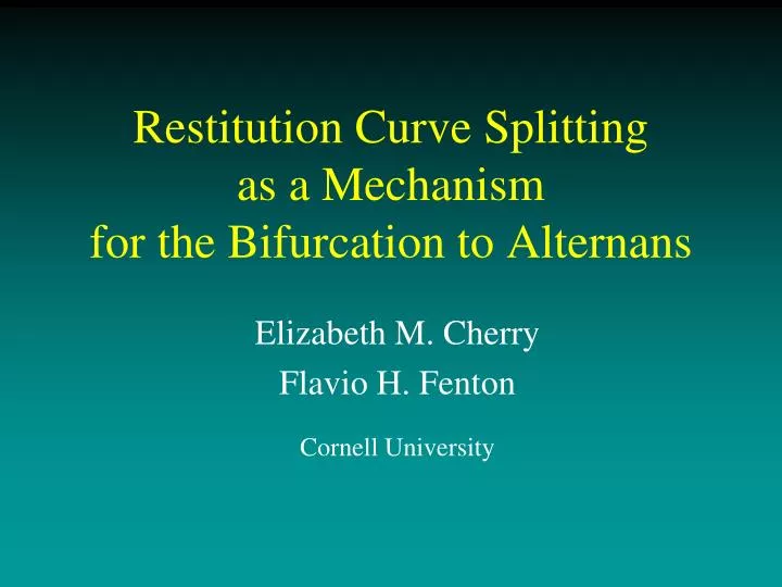
Restitution Curve Splitting as a Mechanism for the Bifurcation to Alternans. Elizabeth M. Cherry Flavio H. Fenton Cornell University. Alternans. Alternans is a beat-to-beat alternation in cellular action potential shape and duration that leads to alternans in the T-wave of the ECG.

E N D
Restitution Curve Splitting as a Mechanism for the Bifurcation to Alternans Elizabeth M. Cherry Flavio H. Fenton Cornell University
Alternans • Alternans is a beat-to-beat alternation in cellular action potential shape and duration that leads to alternans in the T-wave of the ECG. • Alternans can be electrical and/or mechanical; here we focus on electrical. Alternans often precedes more dangerous arrhythmias like ventricular fibrillation. Wellens HJ. 1972. Chest 62, 319. Raeder RA, et al. 1992. N Engl J Med 326, 271.
Alternans • At the cellular level, alternans appears as a beat-to-beat long (L)-short (S) alternation despite a constant pacing period (cycle length CL). • Action potential duration (APD) and/or amplitude may alternate.
Alternans Bifurcation • Plotting APD as a function of cycle length (period) shows a period-doubling bifurcation. • At short cycle lengths, the curve usually terminates when every other beat is blocked.
DI APD APD Restitution APD (ms) Restitution Curve • Plotting APD as a function of preceding DI gives the restitution curve, which provides a first approximation of the system’s dynamics. • Defining APDi+1=f(DIi)=f(CL-APDi) gives an iterative map (for a fixed period CL=APDi+DIi). • Linearizing around the fixed point APD*=f(DI*) by letting APDi=APD* + APDi gives APDi+1=-f’(DI) APDi , which has a bifurcation when |f’(DI)| = 1.
APD Restitution Slope=1 APD (ms) Restitution Curve • APDi+1=f(DIi) is stable when |f’(DIi)|<1. • Alternans is predicted for |f’(DIi)|<1. Cobwebbing technique useful for visualization of the iterative map. (Similar to logistic map.) JB Nolasco, RW Dahlen. 1968. J Appl Physiol 25, 191. MR Guevara et al. 1984. Comput Cardiol, 167. A Karma. 1993. Phys Rev Lett 71, 1103.
APD Restitution Slope=1 APD (ms) Restitution Curve • APDi+1=f(DIi) is stable when |f’(DIi)|<1. • Alternans is predicted for |f’(DIi)|<1. JB Nolasco, RW Dahlen. 1968. J Appl Physiol 25, 191. MR Guevara et al. 1984. Comput Cardiol, 167. A Karma. 1993. Phys Rev Lett 71, 1103.
APD (ms) APD (ms) DI (ms) Period (ms) Alternans Characteristics In models and one-dimensional maps, alternans is characterized by nested “boxes” and large magnitude. But in experiments, alternans boxes slide down and the magnitude of alternans is smaller.
Restitution Curve Splitting • Sliding boxes can be explained if the restitution curve splits into two branches. • Each DI,APD pair then involves one point from each branch. • Branches can have any slope, steep or flat. Canine Purkinje fiber
Restitution Splitting in Experiments • Microelectrode recordings are used. • Decreasing temperature can increase alternans and splitting.
Restitution Splitting in Experiments Slope 1 • During alternans, lines with slope one connect DI,APD pairs. • Branch slopes can be steep or flat. • Purkinje fibers more likely to be steep; epicardium and endocardium more likely to be flat. • “Long” branch can be above or below “short” branch. STEEP FLAT FLAT FLAT
Calcium in Experimental Alternans • Reducing extracellular Ca2+ (from ~2 mM to 62.5M) removes splitting but also eliminates alternans.
APD (ms) APD (ms) CL (ms) DI (ms) 2-Dimensional Map Model • A 2-dimensional map model using both DI and peak calcium to predict APD can reproduce splitting: APDn+1 = f (DIn,Can+1). • Purkinje splitting data are well-matched.
Restitution Splitting in Models • Some models also exhibit restitution curve splitting. • Branches for FMG model have slope > 1, for HR model have slope < 0.5. Fox JJ et al. 2002. Am J Physiol 282, H516. Hund T, Rudy Y. 2004. Circulation 110, 3168. Cherry EM and Fenton FH. 2007. Am J Physiol 292, H43.
Enhancing Restitution Splitting • In the FMG model, restitution splitting can be enhanced by decreasing the exponent in the Jup term (0.5-2.0). Exponent decreasing
Summary • Restitution splitting occurs experimentally in normal canine Purkinje fibers and ventricular tissue. • Slope of restitution curve branches can be steep or flat; no slope > 1 criterion for alternans. • Models, 2-d map also can exhibit splitting. Acknowledgements: Robert F. Gilmour, Jr., NSF, NIH Web site: http://thevirtualheart.org