Download
1 / 24
240 likes | 358 Vues
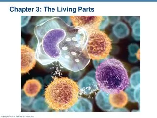
Living Parts. Prokaryotes, Eukaryotes Tissue – groups of cells together for certain specialized functions, differentiated cells. Tissue – 14 major types of tissues in animals epithelial, connective, nervous, muscle, etc. http://www.emc.maricopa.edu/faculty/farabee/BIOBK/BioBookAnimalTS.html.

E N D
Living Parts • Prokaryotes, Eukaryotes • Tissue – groups of cells together for certain specialized functions, differentiated cells • Tissue – 14 major types of tissues in animals • epithelial, connective, nervous, muscle, etc. • http://www.emc.maricopa.edu/faculty/farabee/BIOBK/BioBookAnimalTS.html • Cell – over 200 types in a vertebrate Long – eg. nerve cells Some do not divide for ~ 100 years Some divide rapidly, ~ few hours
Components of a Cell (Eukaryotes) ~70% water 4% small molecules 15-20% proteins 2-7% DNA/RNA 4-7% membrane Picture from on-line biology book, http://www.emc.maricopa.edu/faculty/farabee/BIOBK/BioBookCELL2.html
Membrane • Lipid bi-layer Phospholipids and other lipids hydrophilic, hydrophobic • Small molecules and membrane-bond proteins • Semi-permeable / Osmosis • N2, O2, water, glycerol, glucose, sucrose, Ions, etc. • http://en.wikipedia.org/wiki/Cell_membrane • Picture from : • http://www.cbc.umn.edu/~mwd/cell_www/chapter2/membrane.html
Cytoplasm • Cytoskeleton – fibrous protein complexes • maintain shape, anchoring, moving • actin filaments • microtubules • Ribosome – protein synthesis • Mitochondrion – energy • Endoplasmic reticulum (ER) – mesh of membrane, protein synthesis and transport • Lysosomes, Golgi, vesicles etc. • A good reference sitehttp://www.emc.maricopa.edu/faculty/farabee/BIOBK/BioBookCELL2.html
Nucleus • Nuclear membrane • Nuclear envelope with pores • DNA/RNA and some proteins • A good reference sitehttp://www.emc.maricopa.edu/faculty/farabee/BIOBK/BioBookCELL2.html
Nucleic Acids • DNA – polymers of deoxyribonucleic acids, ds • Nucleotide: 3 components: base (purine/pyrimidine) sugar (ribose/deoxyribose) phosphate group A (adenine) G (guanine) C (cytosine) T (thymine, DNA) U (uracil, RNA) RNA: in both nucleus and cytoplasm, ss 3 types: mRNA, rRNA and tRNA • Picture from on-line biology book • http://www.emc.maricopa.edu/faculty/farabee/BIOBK/BioBookCHEM2.html
Protein-a chemical view • A chain of amino acids folded in 3D • Peptide • Protein backbone • N / C terminal • Picture from on-line biology book
Amino Acids • Different properties – side chain • 20 types in nature Generally: • Positively charged – Arg, His, Lys • Negatively charged – Asp, Glu • Polar but uncharged – Ser, Thr (OH), Asn, Gln(CO) • Special – Cys, Gly, Pro • Hydrophobic – Ala, IIe, Leu, Met, Phe, Trp, Tyr, Val, • A good reference sitehttp://www.emc.maricopa.edu/faculty/farabee/BIOBK/BioBookCELL2.html
Protein – a 3D view • Bond length, bond angle – fairly restricted • Torsion angles on backbone • (phi), (psi), (omega) • , mostly plane(180°, rare case 10°in cis) • , , free but with an average characteristic distribution- Ramachandran plot • Picture from http://www.expasy.org/swissmod/course/text/chapter1.htm
Torsion Angles • Dihedral angles (phi), (psi), (omega) N C C N
Secondary structures • Helix - hydrogen bond (CO)i-(NH)i+4 -helix (3.613) 1.5Å / residue • -sheet is composed of multiple -strands • Hydrogen bond between two -strands • Zig-zag backbone, side-chains opposite directions , ~30°/residue twist, mostly antiparallel • Turn, loop/coil Picture from www.expasy.org site http://www.expasy.org/swissmod/course/text/chapter1.htm
Protein tertiary and quaternary structure • Tertiary – 3D folding of a polypeptide chain involves non-local interaction • Quaternary – multiple chains/multi subunits PDB: http://www.pdb.org SCOP database – protein classification
From DNA to Protein • Genome, genes, chromosome, proteome • Overview of HGP Picture from doegenomics.org http://www.ornl.gov/TechResources/ Human_Genome/project/info.html • Transcription (DNA-mRNA) • Translation (mRNA-polypeptide) • - Gene expression
Transcription • Initiation, Elongation and Termination • Central enzyme: RNA polymerase Picture from http://edtech.clas.pdx.edu/gene_expression_tutorial/transcription.html • RNA polymerase bind to promoter site, e.g. in bacteria 35 BP upstream of start: RNA polymerase binding site (TTGACA) 10 BP upstream of start: box (TATAAT) - sigma factor site • Promoter sequence determines transcription level
Transcription in Eukaryotes • More complicated process • RNA Splicing – intron and exon Picture from http://www.intouchlive.com/home/frames.htm?http://www.intouchlive.com/cancergenetics/genefx.htm&3 • Alternative splicing – diversity of proteins
Translation • Genetic coding • What is a codon? • Ribosome bind upstream region • of mRNA • tRNA bind to specific amino acid • (AUG) on mRNA to start • tRNA brings a.a. to ribosome • At least one tRNA exists for each amino acid Example of a tRNAhttp://users.rcn.com/jkimball.ma.ultranet/BiologyPages/T/Translation.html Picture from http://edtech.clas.pdx.edu/gene_expression_tutorial/translation.html
Regulation in gene expression • Various needs for gene expression • Spatially and timely different steps in eukaryotes • Prokaryote – e.g. lac gene regulation http://users.rcn.com/jkimball.ma.ultranet/BiologyPages/L/LacOperon.html • Eukaryotes Altering rate of transcription Rate of transcript processing, stability of mRNA, efficiency of ribosome • Basel promoter, upstream promoter • Enhancer, silencer • Transcription factors http://users.rcn.com/jkimball.ma.ultranet/BiologyPages/P/Promoter.html
Experimental techniques • Identify size of protein/DNA • e.g. gel electrophoreses • Identify proteins • e.g. using antibodies - structural • Sequencing peptide • e.g. mass spectrometry • Sequencing DNA/RNA • Determine some 3D protein structure • Molecular cloning, producing large amount of genes and proteins
Recombinant DNA technology • Restriction enzyme, ligase • Vector – plasmid, bacteriophage (virus)
Recombinant DNA technology • Cleave DNA • Vector to carry DNA for cloning • Transform bacteria • An example • Grow bacteria • http://www.biology.arizona.edu/molecular_bio/problem_sets/Recombinant_DNA_Technology/05t.html • Screen for cloned DNA • Revolutionized biology
Related techniques • cDNA, vs. genomic DNA • reverse transcriptase • represent currently active mRNA population • function, stage of the cell A cool animation http://www.maxanim.com/genetics/cDNA/cDNA.htm • Polymerase Chain Reaction (PCR) • in-vitro amplification of a region of DNA with known sequence • primer, template • DNA polymerase • http://en.wikipedia.org/wiki/Polymerase_chain_reaction
Protein Structure Determination • X-ray crystallography • soluble, medium size, some viruses • usually difficult for large proteins • Nuclear Magnetic Resonance (NMR) • small , multi-dimensional NMR • Other developing methods • e.g. electron cryomicroscopy • Structural genomics
Protein Crystals Diffraction data Phase Structure Electron density map Sequence X-ray crystallography X-ray Grow suitable crystals – tricky Solving structure – mostly a mature technique
Electron cryo-microscopy • 2D crystallography – e.g. membrane proteins • Non-crystalline • – e.g. viruses, large complexes, helical objects • Take 2D images using TEM • Computationally build 3D structure • Computationally more intensive http://en.wikipedia.org/wiki/Cryo-electron_microscopy