Download

1 / 17
170 likes | 270 Vues
Beauty. Reference 1. C 4. Fe 3+ O x (OH) y core. One bundle. C 3. C 4. Fe 3+ O(OH). Fe 2+ oxid. Fe 2+ exit. 24 subunuits of 4 a-helix bundles. Like an iron malted milk ball!. C 3. At pH 7, [Fe 3+ ] = 10 -18 M

E N D
C4 Fe3+Ox(OH)y core One bundle C3 C4 Fe3+O(OH) Fe2+oxid Fe2+exit 24 subunuits of 4 a-helix bundles Like an iron malted milk ball! C3
At pH 7, [Fe3+] = 10-18 M Ferritin manages to concentrate ferric ion to mM concentrations (10-3 M). Lack of gene for ferritin lethal. The reaction: 2000 Fe2+(H2O)6 + O2 1000 Fe3+—O-O—Fe3+ 2000 Fe3+(H2O)6+ H2O2 fast, ms slow, min or hrs Enters at C3 pores; in subunits ‘translocating mineral precursor’ Fe3+2O3 (H2O)1000 + 5000 H+ Exits from C3 pores
ferroxidase Ferroxidase in H subunits (H for heart, NOT heavy!)
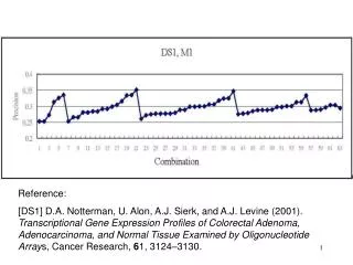
How and where iron exits from ferritin for cellular use is uncertain. Proline substitution for conserved leucine 134 (L134P) allowed normal assembly but increased iron exit rates. X-ray crystallography of H-L134P ferritin revealed localized unfolding at the 3-fold axis, also iron entry sites, consistent with shared use sites for iron exit and entry. The junction of three ferritin subunits appears to be a dynamic aperture with a "shutter" that cytoplasmic factors might open or close to regulate iron release in vivo. Localized unfolding at the junction of three ferritin subunits. A mechanism for iron release? Takagi, H., Shi, D., Ha, Y., Allewell, N.M., Theil, E.C. (1998) J.Biol.Chem. 273: 18685-18688
Someone let the organometallic chemists out … and look what happened.. Apo-ferritin (apo-Fr) mutants are used as scaffolds to accommodate palladium (allyl) complexes. Various coordination arrangements of the Pd complexes are achieved by adjusting the positions of cysteine and histidine residues on the interior surface of the apo-Fr cage. ..


![Reference: [1] TeamSpace paper](https://cdn2.slideserve.com/3924279/reference-1-teamspace-paper-dt.jpg)


![[Reference]](https://cdn3.slideserve.com/6885026/reference-dt.jpg)


![Reference: [1]](https://cdn5.slideserve.com/9521244/5-sources-of-errors-5-2-noise-types-5-2-1-thermal-dt.jpg)