Download
1 / 55
550 likes | 988 Vues
Anemias. 1. In general 2. Decreased erythrocyte production 3. Increased erythrocyte loss. Anemias 1. Anemias in general Definition - traditional RBC or Hb or HTC - alternative Erythron (M)

E N D
Anemias • 1. In general • 2. Decreased erythrocyte production • 3. Increased erythrocyte loss
Anemias 1. Anemias in general Definition - traditional RBC or Hb or HTC - alternative Erythron (M) M = I * T, where I = amount of new RBC produced per unit of time T = red blood cell life span Example (extreme compensation): M = 8 * 1/8 = 100%
Symptoms: under 80g Hb/L Hemolysis jaundice, splenomegaly, cholelithiasis O2 diffusion vasoconstriction of skin and kidneys Pulmonary and cardiac function Medullary erythropoiesis 2,3 diphosphoglycerate shift of the Hb curve to the right O2 delivery to the tissues Acute blood loss: 30% of volume (1500 mL) circulatory colaps, shock 50% loss death Hb after 2-3 days No “emergency” pool of RBC, premature release of reticulocytes only The marrow RBC production can rise up to 8times, if there is Fe enough
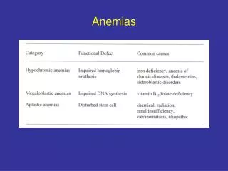
2 Decreased erythrocyte production 21 Decreased proliferation of new erythrocytes = aplasticanemia s.l. = hypoproliferative anemia (Fig. 15) 15
Reticulocyte index . Hypoplasia of the red cell line in the marrow inability to react to anemia • Name: in fact, the anemia is hypoplastic only (never complete aplasia) • Symptomatology: pancytopenia always present (white cells, platelets), infections, bleeding, reticulocytes, plasma Fe, total binding capacity • Prognosis not very good. Ther.: bone marrow transplantations and immunosuppression (cyclosporine and antilymphocyte serum)
Etiology • - “idiopathic” – most often, probably caused by so far • unknown pollutants • known causes - primary (= inborn) – Fanconi´s - secondary (= acquiered): • 211 Decreased erythropoietin • Impaired production by the kidneys - anemia of • renal failure • Low oxygen requirements - anemia of endocrine • disease (hypothyreoidism) • Impaired stem cell response to erythropoietin - • anemia of chronic diseases (see later)
212 Bone marrow damage or defect • Replacement of marrow by tumor (crowding out) • - myelophthisic anemia • Replacement of normal marrow by cancerous • cell line - anemia associated with • myeloproliferative disease • Local competition for nutrients, secretion of • inhibitory substances • Damage to bone marrow by physical or chemical • agents, or infections – aplastic anemia s.s. • Benzene, chloramphenicol, analgesics, • anticonvulsants, antianxiety drugs • Inherited bone marrow defect - Fanconi's • anemia: multiple congenital abnormalities, • recessive gene
22 Impairment in the maturation of new erythrocytes ineffective erythropoiesis Subcellular pathology defective erythroblasts intramedullary hemolysis (>50%). The marrow is hypercellular, in spite of this, the reticulocytes are scanty, however ineffective erythropoiesis 221 Macrocytic-normochromic erythrocytes
Folic acid deficiency (Megaloblastic anemia, Fig. 16) Folate compounds widely distributed in nature, rich in the dietbut small body stores. 16
Folate compounds widely distributed in nature, rich • in the diet, but small body stores. Fig. 17. 17
Decreased intake: alcoholism, hepatic diseases, • tropical sprue (coliform bacteria), malabsorption, • resections. Increased requirements: gravidity, growth, • hematopoiesis. Folic acid antagonists: cytostatics, • chemotherapeutics, antiparasitic and anticonvulsive • drugs • Function of tetrahydrofolate: coenzyme for single- • carbon transfers necessary for thymidylate • synthase rate limiting for DNA synthesis • Vitamin Bl2 deficiency (Megaloblastic anemia) • Synthetized by microbes only in foods of animal • origin. Requirements small, stores large. • Absorption of B12 Fig. 18
Function of B12: myelin (funicular myelosis) and folate synthesis • Deficiency: • - total vegetarianism • - malabsorption syndromes (jejunal bacterial • overgrowth, enteritis, intestinal parasites) • - lack of intrinsic factor pernicious anemia • adults: genetic, autoantibodies against • parietal cells or IF chronic • atrophic gastritis • children: rare, inherited, abnormalities of IF
222 Microcytic-hypochromic erythrocytes Iron deficiency (Iron deficiency anemia) • Absorption is regulated by the needs (i.e., by hematopoiesis) – by the Fe content in the mucosal gut cells • Etiology of Fe deficiency: blood losses, need, malabsorption • Iron metabolism (Fig. 19)
Incorporation of Fe into the erythroblasts only from transferrin Fig. 20 20
Ferritin: a protein envelope surrounds a microcrystalic • core of inorg. Fe • Fe overload: disturbance of the gut mucosal cells. • Hemosiderosis= RES cell containing hemosiderin, • hemochromatosis = various organs contain • hemosiderin • Clinic: angular stomatitis, glossitis, koilonychia, • dysphagia, pica, no sideroblasts, typical serum • composition (Fig. 21)
Other microcytic-hypochromic anemias • Abnormalities of the heme or globin synthesis • Hb production more than 4 divisions • microcytosis and hypochromia • Unavailability of iron to blast cells - Anemia of • chronic disease (ACD) • Block of Fe metabolism: macrophages • degrading Hb of the decayed RBC are activated, • proliferate and retain Fe for themselves • hepatic syntheses transferrin together with • plasma Fe • (Fig. 23)
Fe storage normal or • Besides • - EPO production or its binding to the stem • cells (loss of EPO receptors, disturbed • coupling) • - RBC life span • Etiopathogenesis: foreign antigenes pertaining to • the inflammatory process activation of • macrophages • Il-1 and TNF production: Fe metabolism in • MF disturbed; direct inhibition of EPO • production • Conditions: chronic inflammations (e.g. hepatitis), • some malignancies, collagen-vascular diseases
Impairment of heme synthesis (Sideroblastic = sidero- • achrestic anemia) • Fe into mitochondrias ring erythroblasts • Etiology: • inherited – aminolevulate synthetase – rate limiting • acquiered – somatic mutation – cell clone Impairment of globin synthesis (Thalassemia syndromes) Fig. 24
3 Increased erythrocyte loss • 31 Hemorrhage hemorrhagic anemia • Acute: influx of interstitial fluid into the circulation • (several days) progressive fall of Hb, • HTC, RBC • EPO and reticulocyte response (Fig. 25 and 26) • Chronic: GI ulcers and malignancies, menstruation. • Iron stores
32 Intravascular hemolysis or premature phagocyto- • sishemolytic anemia • 321 Hereditary factors • Defects in the erythrocyte membrane • Hereditary spherocytosis • RBC flexibility is conditioned by the • unique structure of the RBC membrane, • and this is maintained by actin, spectrin, • and ankyrin. Spectrin gene mutates most • often • a) Mutated RBC loss of flexibility • pitting in the splenic sinuses RBC • shrinking getting spherical loss of • flexibility destruction in the spleen
b) Mutated RBC slowering of the • Na/K-ATPase Na into RBC water • into RBC getting spherical etc. • c) Mutated RBC enhanced need of • glucose lowered glucose • concentration in the spleen enhanced • trapping of the microspherocytes • Symptomatology: • Mild anemia only • Bilirubin gale stones • Osmotic fragility test - series of salt • solutions of increasing concentration • sensitivity of RBC to a medium • without glucose
Surface counting patterns of 51Cr-labeled RBC (Fig. 28) • Splenectomy! 28
Hereditary elliptocytosis • Mild anemia, in combination with other • anemizing factors only • Again a membrane defect of actin- • spectrin-ankyrin skeleton • No therapy • Defects in erythrocyte metabolism • G6-PD deficiency anemia • 95% of all glucose metabolism enzyme • deficiencies • Triggering factors: oxidizing drugs, • infections ( activation of leucocytes • producing active oxygen radicals) • Favismis a unique phenomenon – fava • beans (contain an oxidant L-dopa)
Measuring of the G6-PD activity in the • RBC changing of the eating habits • Pyruvate kinase deficiency anemia • Embden-Meyerhof pathway, decline of • ATP production, symptoms may be • severe, specific enzyme assays (no other • specific features), drugs not implicated • in pathogenesis • Abnormal hemoglobin production • Point mutations of Hb are mostly • innocent, a small fraction is pathogenic: • solubility and precipitation, • affinity to oxygen, unstability of • quaternal structure and Hb denaturation
Sickle-cell anemia, HbC disease, HbD • disease, HbE disease • See lecture on genetics • 322 Acquired accelerated hemolysis • Approximately: • hereditary hemolysis factors intrinsic to the RBC • acquiered hemolysis factors extrinsic to the RBC • Physiological aging of RBC their defense mechanismsintravascular hemolysis or phagocytosis in mononuclear phagocytes (MF, reticular and endothelial cells). The environmental stressing factors shorten the RBC life span further. They are present to a degree already under physiologic conditions in most people.
These physiological/pathophysiological factors are of • chemical, physical or immunological nature. The • boundary between physiological and pathological is • fuzzy here: • - AB0 incompatibility is present in 23% of all • gestations (hemolysis is very uncommon here, • however) • - Paroxysmal nocturnal hemoglobinuria is rather • common (mild hemolysis) • - Metabolic stress is ubiquitous
3221 Activation of the immune system immuno- hemolytic anemia Classification of immunohemolytic anemia (IHA) Alloantibodies hemolytic disease of the newborn (HDN) AB0 Rh Autoantibodies Warm-active antibodies Cold-active antibodies Alloantibodies AB0 incompatibility
Mother 0 has antibodies against A and B already spontaneously (in contradisticition to Rh- mothers, in whom the antibodies not are formed before the first parturition), and these reach the foetus via placenta. Not easily, however – they are IgM, therefore large molecules, so the symptoms are mild Antigen-antibody complexes complement activation premature lysis of RBC Rh incompatibility– (Fig. 29)
15% of Rh- mothers At the first parturition, the RBC penetrate from the foetus into the mother production of anti-Rh+-antibodies. The RBC having penetrated during the parturition could be killed by timely administration of anti Rh+ antibody (RhoGAM). In the next gravidity, small numbers of the fetal RBC may get into the maternal circulation anamnestic response higher antibody titers of IgG (readily pass the placenta) HDN in the foetus Symptoms: Hemolysis erythroblastosis Inability to conjugate bilirubin jaundice (possibly s.c. kernikterus, into the basal ganglia)
Residual concentration of the mother´s antibody after birth slowering of growth; exchange transfusion • Autoantibodies autoimmune hemolytic anemias (AIHA) • Warm type AIHA • IgG against RBC membrane antigens • Types: • Idiopathic – antibodies against the proper RBC are formed. Common autoimmunity mechanisms could be considered:
- Molecular mimicry with some microbe • - Lowered function of TS production of • antibodies • - Polyclonal B cell activation • - Enhanced presentation of antigens etc. • Drug induced (Fig. 30)
The drug is a hapten and in a complex with a carrier protein production of antibodies. Three possibilities: • - a drug with a surface RBC antigen neoantigen production of antibodies and opsonization, e.g., penicillin • - a drug and serum protein (instead of RBC protein) neoantigen complexes are deposited on RBC membrane, e.g., antimalarics, sulfonamides, phenacetin • Alfa-methyldopa (antihypertensive drug) triggers the gene for the Rh factor • With other diseases: leukemias, SLE, inf. mononukleosis
RBC coating with an antibody = opsonization binding of MF on RBC pitting by means of nipping of RBC sphericity loss of flexibility trapping Opsonization rarely leads to intravascular hemolysis Symptoms of warm-type AIHA: Are mild, hemolysis is compensated Pitting spherocytes, microcytes Direct Coombs test (Fig. 32): Antiserum against anti-RBC antibodies is formed in rabbits RBC agglutination. IgG alone cannot bridge the repulsive force between RBC (= zeta potential), IgG + anti-IgG antibody can do it agglutination Therapy: corticosteroids, immunosuppresive drugs