Download
1 / 26
280 likes | 806 Vues
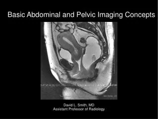
Pelvic floor imaging. Jennifer Kruger 1 Hans Peter Dietz 2 , . 2 Department of Obstetrics and Gynaecology University of Sydney, Sydney, Australia . 1 Department of Sport and Exercise Science, University of Auckland, Auckland, New Zealand . THE UNIVERSITY OF AUCKLAND

E N D
Pelvic floor imaging Jennifer Kruger1 Hans Peter Dietz2, 2Department of Obstetrics and Gynaecology University of Sydney, Sydney, Australia 1Department of Sport and Exercise Science, University of Auckland, Auckland, New Zealand THE UNIVERSITY OF AUCKLAND DEPARTMENT OF SPORT AND EXERCISE SCIENCE
Outline • Pelvic floor imaging in general • 2D imaging • General use of 3D ultrasound • 3D pelvic floor imaging • Pelvic floor function • Research of pelvic floor (pf) function in elite nulliparous athletes using 3D ultrasound. • Possible clinical implications
Introduction • Ultrasound imaging of pelvic floor available for many years • Lack of ionizing radiation • Ease of use • Cost effectiveness • Recently - capable of imaging in multiple planes, 3D images, ‘real time’ property. • These proved useful particularly obstetrics, gynaecology, paediatrics and cardiology I N T R O D U C T I O N THE UNIVERSITY OF AUCKLAND DEPARTMENT OF SPORT AND EXERCISE SCIENCE
2D imaging • Previously 2D/B mode ultrasound scanning used define pathology and normal function of pelvic floor. • Abdominally, intravaginal or transperineal. • Function assessed: • Descent of bladder, uterus and rectal ampulla during a valsalva manouevre. • Images in the mid-sagittal plane. • Still widely used. 2D Imaging THE UNIVERSITY OF AUCKLAND DEPARTMENT OF SPORT AND EXERCISE SCIENCE
2D imaging • Measurements of bladder neck descent and urethral rotation. 2D Imaging Ultrasound images showing measurement of bladder neck descent and urethral rotation. Bladder neck descent (BND)= x-r –x-s. (Dietz et al 2006) THE UNIVERSITY OF AUCKLAND DEPARTMENT OF SPORT AND EXERCISE SCIENCE
3D ultrasound imaging • Use of 3D ultrasound popularised by obstetric scanning. • Volume images of the fetus • Some suggestion improves diagnostic capabilities • Cleft palate • Spinal defects • Gynaecological pathologies • Quantify volumes in urethra and paravaginal supports • Anal canal anatomy and mammography 3D IMAGING THE UNIVERSITY OF AUCKLAND DEPARTMENT OF SPORT AND EXERCISE SCIENCE
3D pelvic floor imaging • Recent advances in 3D/4D transperineal ultrasound imaging suitable for visualisation of pelvic floor muscles. 3D Pelvic floor imaging THE UNIVERSITY OF AUCKLAND DEPARTMENT OF SPORT AND EXERCISE SCIENCE
3D pelvic floor imaging • Display mode on ultrasound machine shows multiplanar images in sagittal, coronal and axial view and a rendered volume image. • Volume image is the integration of 2D sectional images. • Acquisition of multiplanar images allow access to the axial plane – previously domain of magnetic resonance imaging. 3D PELVIC FLOOR IMAGING UNIVERSITY OF AUCKLAND DEPARTMENT OF
3D pelvic floor imaging 3D Pelvic floor imaging A standard acquisition screen of pelvic floor imaging as captured with a Voluson 730 expert system. The orthogonal views are seen at the top left (A plane), top right (B plane), and bottom left (C plane). The bottom right image shows a rendered volume image of the entire levator hiatus. (Dietz et al 2006) UNIVERSITY OF AUCKLAND DEPARTMENT OF
3D pelvic floor imaging – assessing function • Unique plane of acquisition for levator hiatal area: • ‘plane of minimal dimensions’ • Smallest distance from the inferior edge of the symphysis pubis to the anal rectal angle • Levator hiatal area bounded by the symphysis pubis anteriorly, anal rectal angle posteriorly, puborectalis/pubococcygeus laterally. • Hiatal area measures – pelvic floor function • Rest • Maximum pelvic floor muscle contraction • Maximum valsalva 3D PELVIC FLOOR IMAGING THE UNIVERSITY OF AUCKLAND DEPARTMENT OF SPORT AND EXERCISE SCIENCE
3D imaging 3D PELVIC FLOOR IMAGING A mid-sagittal image. Line indicates plane of minimal dimensions B corresponding axial image showing entire levator hiatus ( dotted area) UNIVERSITY OF AUCKLAND DEPARTMENT OF
Protocol for 3D pelvic floor imaging • Transperineal imaging: • GE Kretz Voluson 730 Expert ( similar) • Wide angle of acquisition (85°) curved array volume transducer 8-4MHz. • Imaged supine after voiding • Transducer ‘sits’ on the perineum mid-sagittal orientation • Mid-sagittal/Axial image on the screen • Symphysis pubis reference point – during movement • Evaluation post processing – proprietary software • Methods highly reproducible (Guaderrama 2005, Yang 2006, Dietz 2006 ) 3D PELVIC FLOOR IMAGING THE UNIVERSITY OF AUCKLAND DEPARTMENT OF SPORT AND EXERCISE SCIENCE
Hiatal area on valsalva • Courtesy H.P Dietz 3D PELVIC FLOOR IMAGING THE UNIVERSITY OF AUCKLAND DEPARTMENT OF SPORT AND EXERCISE SCIENCE
‘Ballooning’ of hiatus 3D PELVIC FLOOR IMAGING Courtesy H.P Dietz THE UNIVERSITY OF AUCKLAND DEPARTMENT OF SPORT AND EXERCISE SCIENCE
Pelvic floor muscle contraction 3D PELVIC FLOOR IMAGING THE UNIVERSITY OF AUCKLAND DEPARTMENT OF SPORT AND EXERCISE SCIENCE
THE UNIVERSITY OF AUCKLAND DEPARTMENT OF SPORT AND EXERCISE SCIENCE
Introduction • Preliminary evidence women in long term high impact sport (HIFIT) athletes • Poor progress in labour (Kruger 2006) • Increased incidence of stress incontinence (Bo 2004) • Previous research demonstrated increased cross-sectional area of levator-ani muscle in HIFIT women (Kruger 2006) • Stress incontinence more prevalent in high impact sports (Eliasson 2002, Nygaard 1997 ) • Association between bladder neck descent, hiatal area on valsalva and pelvic floor dysfunction
Aims • Use transperineal 3D ultrasound to characterise the pelvic floor muscle function in HIFIT women and compare it to an age-matched control group. • Investigate pelvic organ descent in both groups during a valsalva manouevre to determine if there is an association with urinary stress incontinence.
Results • All data normally distributed • Mean age 28.5 and 27.6yrs for HIFIT and controls • All asymptomatic for prolapse • Incidence of SUI in the athletes and controls • 3 HIFIT – SUI – only • 1 HIFIT – UI - only • 2 Controls- SUI & UI • Significant differences levator hiatal area on valsalva and BND between groups. HIFIT group higher values for both parameters + increased pubovisceral muscle diameter • None of the other measured parameters different
Results 1=Hifit 2=Control * HIFIT
Conclusions • HIFIT women differ in several aspects of pf function • > Area of levator hiatus on valsalva • No association between hiatal area on valsalva and SUI - was an association with BND. Caution very small numbers • Importance of BN position during raised intra abdominal pressure • Incidence of SUI in HIFIT – ‘overflow phenomenon’ or may be due to • ? Changes in the connective tissue. • Incidence of SUI in athletes in keeping with that of the general population (10%) (Bo 2006)
Possible clinical implications • Pf muscle function determined by ultrasound • May be a method to aid in ‘risk assessment’ for labour. • Better advise women on the likely obstetric impact of long term, high impact sport. • Development of SUI in athletes during competition – etiology unknown but likely to resolve when sport is stopped (Nygaard 1999)