Download
1 / 56
560 likes | 691 Vues
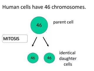
For quite some time, scientists have been interested in chromosomes. WHY???. Chromosomes. They replicate prior to both mitosis and meiosis? How? They carry information for genetic traits (genotype determines phenotype). How?

E N D
For quite some time, scientists have been interested in chromosomes • WHY???
Chromosomes • They replicate prior to both mitosis and meiosis? How? • They carry information for genetic traits (genotype determines phenotype). How? • These are questions of function-to address these questions it seemed logical to look at the structure of chromosomes
Pre-1953-What did we know about chromosomes • What is significant about 1953? • Chromosomes made of DNA and protein • Which of these molecules stored the genetic information? • Most researchers favored protein. Why?
History-DNA or protein is the genetic material? • Griffith-1928 • Avery, McCloud, McCarty-1944 • Hershey and Chase-1952 • Conclusion-DNA was the genetic information in the chromosome • To understand questions of function regarding genes-we had to know the structure of DNA
Mixture of heat-killed S cells and living R cells Heat-killed S cells (control) Living R cells (control) Living S cells (control) LE 16-2 RESULTS Mouse dies Mouse healthy Mouse healthy Mouse dies Living S cells are found in blood sample
Phage head LE 16-3 Tail Tail fiber DNA 100 nm Bacterial cell
The Race to discover the structure of DNA • Watson and Crick • Chargaff • Pauling • Wilkins and Franklin
LE 16-6 Franklin’s X-ray diffraction photograph of DNA Rosalind Franklin
X-ray diffraction insights • Double helix with a uniform width of 2nm • Purine and pyrimidine bases stacked .34 nm apart • Helix makes a turn every 3.4 nm • 10 layers of nitrogen bases every turn of the helix
Purine + purine: too wide LE 16-UN298 Pyrimidine + pyrimidine: too narrow Purine + pyrimidine: width consistent with X-ray data
The “Double Helix” paper • A copy is posted on Angel-please read it • Major insights: • A. DNA is a double helix • B. The two strands are held together by hydrogen bonding between complementary base pairs (A-T) and G-C) • DNA is antiparallel
Sugar–phosphate backbone Nitrogenous bases 5 end Thymine (T) LE 16-5 Adenine (A) Cytosine (C) Phosphate DNA nucleotide Sugar (deoxyribose) 3 end Guanine (G)
LE 16-8a Sugar Sugar Thymine (T) Adenine (A)
LE 16-8b Sugar Sugar Cytosine (C) Guanine (G)
5 end Hydrogen bond 3 end LE 16-7b 3 end 5 end Partial chemical structure
1 nm 3.4 nm LE 16-7a 0.34 nm Key features of DNA structure
LE 16-7c Space-filling model
Structure answers a question of function • Question-How does DNA replicate? • “It has not escaped our notice that the specific pairing we have postulated immediately suggests a possible copying mechanism for the genetic material” • Semi-conservative replication
LE 16-9_1 The parent molecule has two complementary strands of DNA. Each base is paired by hydrogen bonding with its specific partner, A with T and G with C.
LE 16-9_2 The first step in replication is separation of the two DNA strands. The parent molecule has two complementary strands of DNA. Each base is paired by hydrogen bonding with its specific partner, A with T and G with C.
LE 16-9_3 The first step in replication is separation of the two DNA strands. Each parental strand now serves as a template that determines the order of nucleotides along a new, complementary strand. The parent molecule has two complementary strands of DNA. Each base is paired by hydrogen bonding with its specific partner, A with T and G with C.
Experimental Evidence for Semi-conservative Replication • Just because something is logical does not mean it is true. • Three possible mechanisms of DNA replication- • A. Conservative • Semi-conservative • C. Dispersive • Messelson and Stahl experiment
Second replication First replication Parent cell Conservative model. The two parental strands reassociate after acting as templates for new strands, thus restoring the parental double helix. LE 16-10 Semiconservative model. The two strands of the parental molecule separate, and each functions as a template for synthesis of a new, comple-mentary strand. Dispersive model. Each strand of both daughter molecules contains a mixture of old and newly synthesized DNA.
Second replication First replication Parent cell Conservative model. The two parental strands reassociate after acting as templates for new strands, thus restoring the parental double helix. LE 16-10a
Second replication First replication Parent cell Semiconservative model. The two strands of the parental molecule separate, and each functions as a template for synthesis of a new, comple-mentary strand. LE 16-10b
Second replication First replication Parent cell Dispersive model. Each strand of both daughter molecules contains a mixture of old and newly synthesized DNA. LE 16-10c
Figure 16.9 The Meselson-Stahl experiment tested three models of DNA replication (Layer 4)
DNA replication-It’s more complicated than Watson and Crick thought • Considerations-DNA replication • 1. DNA must unwind (it’s a double helix) • 2. It’s fast (mammals-50 nucls/sec; bacteria-500 nucls/sec). • 3.Accuracy-1 mistake/1 billion nucleotides • 4. DNA polymerase limitations-can’t synthesize denovo; only works in 5’3’ direction • 5. DNA is antiparallel
DNA replication proteins • Several of the replication considerations suggest the involvement of proteins (especially enzymes) in DNA replication
Consideration #1-DNA must unwind prior to replication • DNA helicase (unwindase) • Topoisomerase (relieves twisting) • Single strand binding proteins
Consideration #2-Speed of Replication • Enzymes involved-DNA polymerase (11 forms in eukaryotes)-III is the major replicative enzyme) • DNA replication is bi-directional
New strand Template strand 5¢ end 3¢ end 5¢ end 3¢ end Sugar Base LE 16-13 Phosphate DNA polymerase 3¢ end 3¢ end Pyrophosphate Nucleoside triphosphate 5¢ end 5¢ end
Parental (template) strand 0.25 µm Origin of replication Daughter (new) strand LE 16-12 Replication fork Bubble Two daughter DNA molecules In this micrograph, three replication bubbles are visible along the DNA of a cultured Chinese hamster cell (TEM). In eukaryotes, DNA replication begins at may sites along the giant DNA molecule of each chromosome.
Consideration #3-Accuracy • DNA polymerase has “proofreading capabilities”-mismatch repair
Consideration #4-Limitations of DNA polymerase • DNA polymerase can’t synthesize a new strand “denovo”-needs a free 3’ OH group to attach the next nucleotide to • Solution-RNA primase-adds RNA primer (5-10 nucleotides)-later primer removed by a form of DNA polymerase that replaces RNA nucleotides with DNA nucleotides • Pieces of DNA joined by DNA ligase
Primase joins RNA nucleotides into a primer. 3¢ 5¢ 3¢ 5¢ Template strand LE 16-15_1 Overall direction of replication
Primase joins RNA nucleotides into a primer. 3¢ 5¢ 3¢ 5¢ Template strand DNA pol III adds DNA nucleotides to the primer, forming an Okazaki fragment. 3¢ 5¢ RNA primer 3¢ 5¢ LE 16-15_2 Overall direction of replication
Consideration #4-Limitations of DNA polymerase (continued) • DNA polymerase only works in 5’3’ direction • Why is this a problem? • Because of consideration #5-DNA is antiparallel-One strand runs in the 5’3’ direction; the other runs in the 3’5’ direction • Solution1- Is there a 3’5’ DNApolymerase? (haven’t found one yet)
Solution 2-DNA replication occurs differently on the 2 strands • Leading strand (continuous replication) • Lagging strand (discontinuous replication)-involvement of Okasaki fragments (approximately 200 nucleotides in length in eukaryotes).
3¢ 5¢ Parental DNA Leading strand 5¢ 3¢ Okazaki fragments Lagging strand LE 16-14 3¢ 5¢ DNA pol III Template strand Leading strand Lagging strand Template strand DNA ligase Overall direction of replication
Primase joins RNA nucleotides into a primer. 3¢ 5¢ 3¢ 5¢ Template strand LE 16-15_1 Overall direction of replication
Primase joins RNA nucleotides into a primer. 3¢ 5¢ 3¢ 5¢ Template strand DNA pol III adds DNA nucleotides to the primer, forming an Okazaki fragment. 3¢ 5¢ RNA primer 3¢ 5¢ LE 16-15_2 Overall direction of replication
Primase joins RNA nucleotides into a primer. 3¢ 5¢ 3¢ 5¢ Template strand DNA pol III adds DNA nucleotides to the primer, forming an Okazaki fragment. 3¢ 5¢ RNA primer 3¢ 5¢ After reaching the next RNA primer (not shown), DNA pol III falls off. LE 16-15_3 Okazaki fragment 3¢ 3¢ 5¢ 5¢ Overall direction of replication
Primase joins RNA nucleotides into a primer. 3¢ 5¢ 3¢ 5¢ Template strand DNA pol III adds DNA nucleotides to the primer, forming an Okazaki fragment. 3¢ 5¢ RNA primer 3¢ 5¢ After reaching the next RNA primer (not shown), DNA pol III falls off. LE 16-15_4 Okazaki fragment 3¢ 3¢ 5¢ 5¢ After the second fragment is primed, DNA pol III adds DNA nucleotides until it reaches the first primer and falls off. 5¢ 3¢ 3¢ 5¢ Overall direction of replication
Primase joins RNA nucleotides into a primer. 3¢ 5¢ 3¢ 5¢ Template strand DNA pol III adds DNA nucleotides to the primer, forming an Okazaki fragment. 3¢ 5¢ RNA primer 3¢ 5¢ After reaching the next RNA primer (not shown), DNA pol III falls off. LE 16-15_5 Okazaki fragment 3¢ 3¢ 5¢ 5¢ After the second fragment is primed, DNA pol III adds DNA nucleotides until it reaches the first primer and falls off. 5¢ 3¢ 3¢ 5¢ DNA pol I replaces the RNA with DNA, adding to the 3¢ end of fragment 2. 5¢ 3¢ 3¢ 5¢ Overall direction of replication