Download
1 / 36
360 likes | 423 Vues
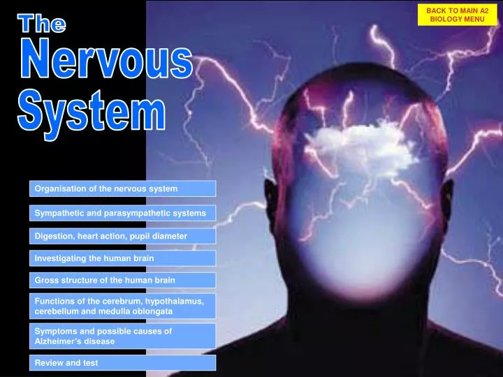
BACK TO MAIN A2 BIOLOGY MENU. The. Nervous. System. Organisation of the nervous system. Sympathetic and parasympathetic systems. Digestion, heart action, pupil diameter. Investigating the human brain. Gross structure of the human brain.

E N D
BACK TO MAIN A2 BIOLOGY MENU The Nervous System Organisation of the nervous system Sympathetic and parasympathetic systems Digestion, heart action, pupil diameter Investigating the human brain Gross structure of the human brain Functions of the cerebrum, hypothalamus, cerebellum and medulla oblongata Symptoms and possible causes of Alzheimer’s disease Review and test
Nervous The The Organisation of the Nervous System System Types of cells Neurones Transmit nerve impulses Help with ionic balance and nutrient supply to the neurones Glial cells e.g. Schwann cells Organisation There are 2 main parts to the nervous system: The Central Nervous System: brain and spinal cord The Peripheral Nervous System: the neurones that lie beyond the brain and spinal cord
Nervous The The Organisation of the Nervous System System The central nervous system - CNS Most neurones in the CNS are intermediate neurones, often each with thousands of synaptic connections These synaptic connections are of 2 types: Excitatory – action potentials in the nerve depolarise the post synaptic membrane of the nerve on the other side of the synapse Inhibitory – action potentials in the nerve hyperpolarise the post synaptic membrane of the nerve on the other side of the synapse There are about 200 x 1012 neurones in the brain and the mix of inhibitory and excitatory synapses provides an almost infinite framework for patterns of nervous activity
The spinal cord The meninges Nervous The The Organisation of the Nervous System System Canal The spinal cord (SC) runs through the neural arches of the vertebrae and in its centre is a canal containing cerebrospinal fluid. Butterfly shaped area of unmyelinated neurones (grey) Myelinated neurones (white) The brain is a highly specialised area of the SC. See later The brain and spinal cord are surrounded by 3 membranes called the meninges The meninges secrete cerebrospinal fluid. The fluid supplies oxygen and nutrients and acts as a shock absorber
Nervous The The Organisation of the Nervous System System Types of cells Neurones Transmit nerve impulses dendrites Node of Ranvier nucleus myelin sheath cell body axon synapse
Nervous The The Organisation of the Nervous System System The peripheral nervous system The cell bodies of sensory neurones are lie just outside the spinal cord in the dorsal root ganglion (a group of nerve cell bodies) The cell bodies of motor neurones are in the spinal cord Axons and dendrites are arranged in bundles called nerves CHECK IT OUT In the knee jerk reflex, the lower leg swings forward quickly when an area just below the knee is tapped. In order for this reflex to occur, a message travels through a sensory neuron to the spinal cord, where an interneuron carries it to a motor neuron, which sends the information to the leg muscle and causes the movement. A typical somatic NS reflex arc
x100 Nervous The The Organisation of the Nervous System System TS Nerve containing 100s of axons and dendrons MYELIN SHEATH AXON Nearly all the nerves shown are myelinated but a few are non-myelinated BACK
CENTRAL NERVOUS SYSTEM (CNS) PERIPHERALNERVOUS SYSTEM (PNS) BRAIN AND SPINAL CORD PERIPHERAL NS SOMATIC NERVOUS SYSTEM (voluntary) AUTONOMIC NERVOUS SYSTEM (involuntary) MOTOR NEURONES TO INTERNAL ORGANS SENSORY AND MOTOR NEURONES TO / FROM SKELETAL MUSCLE SYMPATHETIC NERVOUS SYSTEM (involuntary) PARASYMPATHETIC NERVOUS SYSTEM (involuntary) CONTROLS ORGANS WHEN BODY IS AT REST CONTROLS ORGANS IN TIMES OR STRESS Nervous The The Organisation of the Nervous System System NERVOUS SYSTEM
Series of ganglia Nervous The The Organisation of the Nervous System System The sympathetic nervous system The cell bodies of its motor neurones lie in ganglia outside the spinal cord PUPILS From these ganglia sympathetic motor axons pass to all organs of the body, eventually synapsing with muscles, e.g. cardiac, smooth. SALIVARY GLANDS HEART BRONCHI The transmitter liberated at these synapses is usually nor adrenaline – it stimulates organ activity LIVER STOMACH/ SMALL INTESTINE Ach is released at motor endings in sweat glands, erector muscles and some blood vessels. It too causes stimulation. ADRENAL GLAND / KIDNEYS LARGE INTESTINE BLADDER / GENITALS SNS functions are FIGHT OR FLIGHT
Nervous The The Organisation of the Nervous System System The Parasympathetic Nervous System The nerve pathways all begin in the brain, or at the top or bottom of the SC EYE SALIVARY GLANDS The neurones keep going ‘till right inside the organ. Here they synapse with a motor neurone BRONCHI HEART The transmitter liberated at these synapses is acetylcholine and this has an inhibitory effect on the organ STOMACH PYLORIC SPHINCTER PANCREAS Many parasympathetic axons are part of the vagus nerve. LARGE INTESTINE, ANAL SPHINCTER BLADDER PNS functions are REST AND DIGEST GENITALS
VISCERAL EFFECTOR VISCERAL EFFECTOR Sympathetic fibres VISCERAL EFFECTOR VISCERAL EFFECTOR Nervous The The Organisation of the Nervous System System Layout of motor pathways in the autonomic nervous system Cranial parasympathetic fibres Ach Ach GANGLION NA Ach GANGLION Preganglionic neurone Motor neurone NA Ach Sacral parasympathetic fibres GANGLION Ach Ach
Nervous The The Organisation of the Nervous System System Some sympathetic and parasympathetic effects Effect of sympathetic stimulation Effect of parasympathetic stimulation Organ Increase rate and force of contraction Reduces rate and force of contraction Heart Constricts Eye pupil Dilates Relax – lens thinner for distant vision Contract – lens thicker for near vision ciliary muscles Digestive system glands Little or no effect Stimulates secretion sphincter muscles Contraction Relaxation Small increase in glycogen production liver Release of glucose into blood Little effect, except to increase sweating on palms of hands Increases sweating Skin sweat glands Contract, making hairs stand on end erector muscles No effect vasoconstriction arterioles No effect
Nervous The Sympathetic and parasympathetic action System Effect on the digestive system Salivary glands produce more saliva Reduced blood supply to gastric and salivary glands Peristaltic muscles contract more frequently Reduces peristalsis Sphincter muscles relax Sphincter muscles close Gastric glands secrete more juice PARASYMATHETIC STIMULATES DIGESTION SYMATHETIC WEAKLY SUPPRESSES DIGESTION
Nervous The Sympathetic and parasympathetic action System The action of the heart Ach Ach NA NA PARASYMPATHETIC REDUCES THE RATE AND FORCE OF CONTRACTION SYMPATHETIC INCREASES THE RATE AND FORCE OF CONTRACTION Ach release from the vagus nerve NA release from sympathetic nerves
Nervous The Sympathetic and parasympathetic action System Pupil dilation and constriction
radial circular CLICK TO CONSTRICT PUPIL DILATED Nervous The Sympathetic and parasympathetic action System Pupil dilation and constriction The SNS is stimulating the radial muscles to contract, widening the pupil. The pupil is the dark space in the centre of the iris The iris contains circular and radial muscles and their activity can change pupil diameter Causes: Dim light Excitement Fear
radial circular Nervous The Sympathetic and parasympathetic action System Pupil dilation and constriction CLICK TO DILATE PUPIL CONSTRICTED The PSNS is stimulating the circular muscles to contract, narrowing the pupil diameter The pupil is the dark space in the centre of the iris The iris contains circular and radial muscles and their activity can change pupil diameter Cause: Very bright light
Broca’s area responsible for the production of language THE HUMAN BRAIN Nervous The The Brain System The study of the brain - introduction Difficulties arise because of the enormous number of cells and the huge numbers of connections Thus research often focuses on the function of groups of cells and how they connect to other groups Post-mortem examination of brain tissue from patients with mental disorders was for a long time the only way that brain function could be studied Pierre Broca (France, 1861) for example studied the brain of a patient who could not speak or write but could understand language that he read or heard He found that a small area at the front left side was damaged. Discovery of similar damage in other patients led to this being called Broca’s area.
THE HUMAN BRAIN Nervous The The Brain System The study of the brain – methods used Studies of the brains of people with brain illnesses before or after death, either by using post-mortem brain slices or new scanning methods on living patients. Investigating areas of brain activity in healthy people using new scanning techniques CAT scanning Computer assisted tomography PET scanning Positron emission tomography MRI scanning Magnetic resonance imaging Studying the effect of particular drugs on the brain. Trials with various molecules have provided much information about the structures and functions of molecules found in normal brains
CEREBRUM WITH FOLDED OUTER LAYER CALLED CEREBRAL CORTEX Nervous The The Brain - Structure System EXTERNAL STRUCTURE PARIETAL LOBE FRONTAL LOBE OCCIPITALLOBE TEMPORALLOBE CEREBELLUM MEDULLA OBLONGATA
Nervous The The Brain - Structure System VERTICAL SECTION THROUGH THE HUMAN BRAIN VENTRICLES CONTAINING CEREBROSPINAL FLUID - CSF PARIETAL LOBE FRONTAL LOBE OCCIPITAL LOBE THALAMUS CEREBELLUM MEDULLA OBLONGATA HYPOTHALAMUS PITUITARY GLAND SPINAL CORD
Nervous The The Brain - Function System THE MAJOR PARTS OF THE BRAIN The cerebrum contains the right and left cerebral hemispheres linked by the corpus callosum Thinking, language, planning, emotions, memory The surface of each cerebral hemisphere is covered by a highly folded tissue called the cerebral cortex Control of the autonomic nervous system and some endocrine glands Lobes in the cerebral cortex: frontal, parietal, temporal, occipital. Control and coordination of movement and posture The thalamus and hypothalamus lie below the cerebrum. The hypothalamus is closely associated with the pituitary gland Control of breathing movements, heart rate, action of smooth muscle in the gut
Broca’s area Association areas Primary auditory area Primary visual cortex Nervous The The Brain - Function System The Cerebrum Both cerebral hemispheres receive sensory information from the eyes, ears, skin etc. Areas first receiving this information are called primary sensory areas Areas which then process and integrate this information are called association areas. There are 3 main association areas making up a large part of the cortex. They involve co-ordination such as body position analysis, planning actions and movements, and also creating emotions and memory
Looking at words speaking words Listening to words Thinking of words Nervous The The Brain - Function System The Cerebrum PET scanning of the brain shows that different regions are involved in different stages of a task. E.g. communicating with words In the mid 1800s Paul Broca discovered that 1 small area of the brain was involved in the production of language, speech or writing In 1876Carl Wernicke reported a different are responsible for the understanding of language Although the right and left cerebral hemispheres communicate constantly through the corpus callosum, there is an asymmetrical distribution of sites for language and 3D processing
1824-1880 1848-1904 http://www.uic.edu/depts/mcne/founders/page0013.html http://www.uic.edu/depts/mcne/founders/page0101.html Nervous The System Dr Paul Broca Carl Wernicke
Nervous The The Brain - Function System The right and left cerebral hemispheres
hypothalamus Nervous The The Brain - Function System Functions of the hypothalamus cerebral cortex right thalamus left thalamus cerebellum
Nervous The The Brain - Function System Pituitary Hormones Nerve cells in the hypothalamus synthesise oxytocin and antidiuretic hormone (ADH). HYPOTHALAMUS These travel along axons to the posterior pituitary. They are released from nerve endings into the blood when the nerves are active. Nerve cells in the hypothalamus synthesise and release hormones into the blood stream. This is called neurosecretion. ANTERIOR PITUITARY POSTERIOR PITUITARY The hormones (called releasing hormones) travel to the anterior pituitary where they stimulate further hormone release into the blood (see next table)
Nervous The The Brain - Function System Pituitary Hormones Release of thyroxine from thyroid gland which increases metabolic rate Secretion of thyrotropin releasing hormone Release of thyroid stimulating hormone from the anterior pituitary Secretion of growth hormone releasing hormone Release of growth hormone from the anterior pituitary Growth of cells and tissues FSH: Follicle growth in ovaries. Sperm production in the testes. LH: Ovulation and secretion of oestrogen progesterone and testosterone. Release of LH and FSH hormone from the anterior pituitary Secretion of gonadotrophin releasing hormone Production of antidiuretic hormone (ADH) ADH is released from nerve endings in the posterior pituitary Water retention by the kidney Uterus contraction during birth. Milk ejection during breast feeding Oxytocin is released from nerve endings in the posterior pituitary Production of oxytocin
Nervous The The Brain - Function System Functions of the cerebellum Coordination of movement and posture Does this by smoothing the action of movements initiated by the motor cortex Involved in coordinated task learning – e.g. riding a bicycle, catching a ball Medulla oblongata
Nervous The The Brain - Function System Functions of medulla oblongata Breathing: Controls diaphragm and intercostals via the vagus nerve If blood CO2 changes the medulla can modify the speed and depth of breathing CO2 levels are monitored by receptors in the medulla itself, and also by receptors in the walls of the carotid arteries and aorta. Medulla oblongata Heart rate and blood pressure: Sympathetic and parasympathetic (vagus) nerves link the medulla to the SAN Baroreceptors (pressure receptors) in the carotid arteries and CO2 receptors monitor levels and send information to the medulla High blood pressure or low CO2. Response: Vagus nerve causes the SAN to pulse more slowly Low blood pressure or high CO2. Response: Sympathetic nerves cause the SAN to pulse faster
Alois Alzheimer Nervous The The Brain - Function System Alzheimer’s disease First recorded by Alzheimer after studying the brain of a woman who had died after suffering dementia in 1906. Shrinkage of brain tissue. Ventricles enlarge. short-term memory begins to decline ability to perform routine tasks also declines. Emotional outbursts may occur and language is impaired. Progressively more nerve cells die with subsequent behaviour changes, such as wandering and agitation. ( from the front). The ability to recognize faces and to communicate is completely lost in the final stages. Patients lose bowel and bladder control, and eventually need constant care. The average length of time from diagnosis to death is 4 to 8 years, but can take 20
Nervous The The Brain - Function System Alzheimer’s disease and amyloid plaque formation In Alzheimer's some of the neurones have bundles of fibres in them, called ‘tangles’. Between the neurones there are dark-staining deposits called plaques The tangles in the neurones are made of a protein called tau. The plaques contain a peptide called beta amyloid, Aβ All cell membranes contain a larger protein called amyloid precursor protein or APP. APP to Aβ conversion is part of the normal activity of the cell membrane, but the resulting Aβ is removed by the tissue fluid . Abnormal metabolism of APP may be the cause of some types of Alzheimer’s
Nervous The The Brain - Function System Alzheimer’s Risk factors Ageing. Less than 1 in 1000 people < 65 have Alzheimer's. 1 in 20 > 65 has! A small proportion of sufferers have a genetic (familial) form A varied and active life may help avoid Alzheimer’s. Severe blows to the head (especially in the over 50s) may increase the chance of developing the disease Smoking and high cholesterol may also be risk factors for Alzheimer’s Future therapy? A vaccine to break down the β amyloid plaques? Trials in mice An inhibitor of the membrane enzyme that breaks down APP into Aβ?
Nervous The WHAT YOU SHOULD KNOW AT THE END OF THIS UNIT System REVIEW Describe the organisation of the nervous system with reference to the central and peripheral systems Outline the organisation of the autonomic nervous system into a parasympathetic and a sympathetic system Outline the roles of the autonomic nervous system in controlling the digestive system, heart action and the size of the pupil in the eye Describe the gross structure of the human brain Outline the functions of the cerebrum, hypothalamus, cerebellum and medulla oblongata Describe the symptoms and possible causes of Alzheimer’s disease as an example of brain malfunction. TEST QUESTIONS
Nervous TEST Name ________________ The System Name the 2 major parts of the nervous system 2 Q1 The central and peripheral nervous systems What are the 2 divisions of the peripheral nervous system? 2 Q2 The somatic and the autonomic nervous systems Give 2 features of the parasympathetic stimulation of digestion 2 Q3 Salivary secretion increases, peristaltic increase, sphincters relax, gastric glands secrete more Give 2 features of the sympathetic control of the heart beat 2 Q4 Increased force and increased rate of contraction Outline the effect of the PSNS on light entry into the eye. 2 Q5 Stimulates circular muscles to contract, narrowing the pupil diameter. Name 2 cerebrum functions listed in the table you were given. 2 Q6 Thinking, planning, language, emotions, memory Q7 What is neurosecretion? 3 Synthesis and release into the blood of hormones by nerve sells in the hypothalamus Q8 What are the main function of the cerebellum. Give 1 example of this function. 2 Coordination / smoothing of movement and posture. e.g. flying / riding a bike Q9 The medulla oblongata controls breathing. Name 2 places where CO2 receptors are found 2 In the medulla, in the walls of the carotid arteries and aorta. For what reasons have Paul Broca and Carl Wernike become famous? 2 Q10 ‘Discovered’ centres for: Production of speech, writing. Understanding of language. Q11 Give the 2 most obvious anatomical symptoms of Alzheimer’s disease 2 Shrinkage of brain tissue, enlargement of the ventricles, amyloid plaque formation TOTAL /23