Download
1 / 12
130 likes | 479 Vues
Treatment of Keratoconus Using Riboflavin-Induced Corneal Collagen Crosslinking. Authors: Anne Keating, M.D. 1,2 , Kathryn Colby, M.D., Ph.D. 2 , Roberto Pineda, M.D. 2 , Michael Endl, M.D. 4 , Thomas Elmer, M.D. 4 , Sandra Everett, M.D. 1 and James Reidy, M.D. 1

E N D
Treatment of Keratoconus Using Riboflavin-Induced Corneal Collagen Crosslinking Authors: Anne Keating, M.D.1,2, Kathryn Colby, M.D., Ph.D.2, Roberto Pineda, M.D.2, Michael Endl, M.D.4, Thomas Elmer, M.D.4, Sandra Everett, M.D.1 and James Reidy, M.D.1 1SUNY Buffalo, 2Massachusetts Eye and Ear Infirmary, 3Fichte Endl Elmer Eyecare, Buffalo, NY Financial Disclosure: The authors of this poster have no financial interest in the subject matter of this poster.
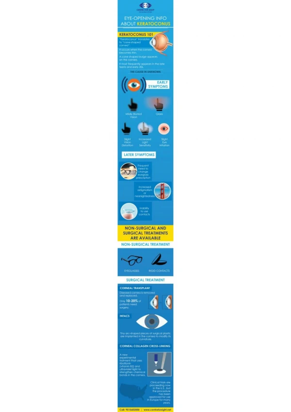
Introduction • Keratoconus is a bilateral disease of corneal collagen fibers that leads to thinning of the cornea and protrusion of the anterior portion of the cornea. The disease affects approximately one in 2000 people of all genders and ethnic groups. The thinning and protrusion of the cornea can lead to tears in the cornea and acute hydrops, where there is sudden corneal edema and eventual scarring and loss of vision. Even without this complication, patients with keratoconus often have decreased vision due to abnormally high amounts of astigmatism, or irregularly shaped corneas. Approximately 20% of people affected by keratoconus will require at least one corneal transplant during their lifetime. The aim of our treatment is to strengthen the corneas by cross-linking the collagen via application of riboflavin and stimulation by UV light so that they do not slowly weaken, reduce vision, cause scarring, and possibly require corneal transplantation. The expectation is that treatment will slow the progression of the disease and avoid the need for a corneal transplant. The treatment has been performed in Europe for the past decade with remarkably favorable results (see Table 1 and references).
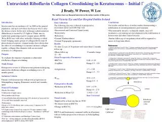
Methods The Caporossi-Baiocchi-Mazzotta (CBM) VEGA X-linker was developed by Aldo Caporossi, Cosimo Mazzotta, and Stefano Baiocchi in collaboratioin with the Italian firm C.S.O. The CBM X-linker is a device that emits ultraviolet (UV) radiation at 370 nm (peak absorption of riboflavin) (figure 1). The device is designed to produce a timely, homogeneous dose of irradiation and deliver it to a 9 mm diameter spot size at a distance of between 1.5 and 1.8 cm. This working distance allows for more efficient focusing on the cornea. A small digital video camera is included in the center of the UV-A array in order to monitor the aiming-beam alignment and to control the centration of the irradiated area. The picture from the video camera is shown on an LCD (liquid crystal display) monitor mounted on the control unit of the equipment (figure 2).
Figure 1 – figure representing the theory of cross linking induced by UVA light and riboflavin
Protocol – slide 1 • The goal is for two groups of approximately 66 patients to be included in the study. One eye from the first group will undergo treatment, while the second group of age matched individuals will serve as a control. This is a prospective, randomized, blinded study. Each patient will be randomized at the beginning of enrollment in the study and will receive a randomization number.
Protocol – slide 2 • The treated and control groups will undergo the following: Two days prior to the procedure, patients will start taking 600 mg of ibuprofen twice daily, which they will continue for a total of 5 days. A drop of 1% pilocarpine will be placed into the treated eye. Topical anesthesia (2% lidocaine jelly) will then be applied and an eyelid speculum will be placed to keep the eyelids open. The central 8.5-9 mm of corneal epithelium will be removed cautiously with the Amoils Epithelial Scrubbe. Riboflavin 0.1% solution will be applied (10 mg riboflavin-5-phosphate in 10 ml dextran T-500 20% solution, supplied in a sterile, single dose container- figure 2) to the cornea every 2-3 minutes for 15 minutes prior to beginning the UV light. The UV source will be from the CBM VEGA X-linker (CSO, Florence, Italy). A wavelength of 370 nm will be used to direct 5.4 J/cm2 to the area of cornea that was debrided for 30 minutes. Every 5 minutes, the UV light will pause briefly while another drop of riboflavin is applied. The distance from the UV source to the cornea will be 1.5 to 5.4 cm. After treatment, the cornea will be rinsed with chilled saline and 2-4 drops of moxifloxacin (Vigamox™, Alcon, Fort Worth, TX) will be instilled, along with 1-2 drops of cyclopentolate 1% (Cyclogel™, Alcon, Fort Worth, TX). A bandage contact lens (FOCUS, 8.4 BC) will be placed prior to sending the patient home and this will be removed after complete re-epitheliaization has occurred, typically within 5 to 7 days. The patients will be asked to use the moxifloxacin four times a day and Acular LS four times a day until the 1 week post-procedure visit (at which time the epithelium will be examined for healing).
INCLUSION CRITERIA for participation: • a. age between 16-35 • b. no prior history of ocular surgery • c. treatment eye must have a corneal power (K power) of between 47 D and 60 D (a measurement of the steepness of the cornea) in the steepest meridian • d. corneal thickness must be greater than 400 µm • d. absence of corneal scarring in the eye to be treated • e. patients must meet the diagnostic criteria for keratoconus, which include one or more of the following features: • -high myopia • -corneal ectasia as viewed by slit-lamp exam or measured by pachymetry • -Vogt’s striae (fine, parallel striations in the corneal stroma) • -topographic findings of superior flattening and inferior steepening of the cornea with 3 or more diopters of difference (see Figure 4) • -presence of Fleischer ring
STUDY DESIGN/MEASURMENTS: Best Corrected Visual Acuity • Corneal Topography as measured by Pentacam • Corneal pachymetry (both by ultrasound and pentacam) • Corneal hysteresis as measured by Ocular Response Analyzer • Intraocular Pressure measured by tonopen • Endothelial Cell Count
Results/Discussion: The treatment of keratoconus using UVA induced riboflavin corneal collagen cross linking has been conducted in Germany and Italy over the past decade with fairly consistent and positive results. The table below summarizes the follow-up and results.