Download
1 / 38
530 likes | 1.14k Vues
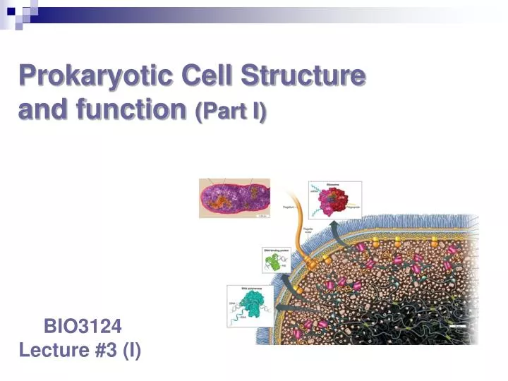
Prokaryotic Cell Structure and function (Part I). BIO3124 Lecture #3 (I). Plasma Membrane Properties and Functions. defines the existence of a cell Made of lipid bilayer Double layer of phospholipids Surrounds the cell approx . 5-10 nm in thickness

E N D
Prokaryotic Cell Structure and function (Part I) BIO3124 Lecture #3 (I)
Plasma Membrane Properties and Functions • defines the existence of a cell • Made of lipid bilayer • Double layer of phospholipids • Surrounds the cell • approx. 5-10 nm in thickness • Separatesexteriorenvironmentfrominterior • Dynamicselectivebarrier • Concentrates certain components intracellulary • Allowsexcretion of waste • Sense the outside world • Severalmetabolicprocesses • ex. Respiration, photosynthesis
Fluid Mosaic Model of Membrane Structure • Lipid bilayer in which proteins float (Singer and Nicholson model)
Membrane proteins Membrane proteins serve numerous functions, including: - Structural support - Detection of environmental signals - Secretion of virulence factors and communication signals - Ion transport and energy storage Have hydrophilic and hydrophobic regions that lock the protein in the membrane
Membrane lipids • Amphipathic phospholipids • polar ends (hydrophilic) • Glycerol, negative charge (outer leaflet) • Ethanolamine, positive charge (inner leaflet) • nonpolar ends (hydrophobic) • Tails of fatty acids • Palmitic acid • Oleic acid (kinking) increase fluidity • Cyclopropane conversion • aging cells Phosphatidylethanolamine
Bacterial Membranes • differ from eukaryotes in lacking sterols • do contain hopanoids, sterol-like molecules • synthesized from similar precursors • Stabilize bacterial membranes • total mass on earth ~1012 tons • a highly organized, asymmetric structure, flexible and dynamic
Archaeal membranes • Etherglycerol, not ester bond • Terpene derived lipids • some have a monolayer membranes • Tetra-ether glycerol • Cyclopentane: isoprene cyclization • Increased stability
Archaeal membranes Moderately thermophilic - Bilayer or mixed Extreme thermophiles eg. Solfolobus and Theromoplasma
Role of cell membrane in energy metabolism • bacterial cell membranes involved in ETC • Gradual energy release • forming proton gradient across membrane
The transfer of H+ through a proton pump generates an electrochemical gradient of protons, called a proton motive force. The Proton Motive Force - It drives the conversion of ADP to ATP through ATP synthase. - This process is known as the chemiosmotic theory.
Besides ATP synthesis, Dp drives many cell processes including: rotation of flagella, uptake of nutrients, and efflux of toxic drugs PMF Drives Many Cell Functions
ATP synthase mechanism Note: pump also works in reverse to create H+ gradient
Cell Transport • Transporters pass material in/out of cell • Passive transport follows gradient of material • Pumps use energy • ATP or PMF • Move material against their gradient • Passive diffusion lets small molecules into cell
The Bacterial Cell Wall • rigid structure that lies just outside the plasma membrane
Functions of cell wall • provides characteristic shape to cell • protects the cell from osmotic lysis • may also contribute to pathogenicity • very few procaryotes lack cell walls, ie Mycoplasmas
Evidence of protective nature of the cell wall • Lysozyme treatment • Penicillin inhibits peptidoglycan synthesis
Gram negative cell wall • few PG layers, defined Periplasmic space • unique outer membrane, LPS, Braun’s lipoprotein Braun’s
Gram positive cell walls • Multiple PG layers, periplasmic space exposed • Teichoic acid
Peptidoglycan (Murein) Structure • Mesh-like polymer composed of identical subunits • contains N-acetyl glucosamine and N-acetylmuramic acid and several different amino acids • chains of linked peptidoglycansubunits are cross linked by peptides
G- G+ Bacterial cell wall
Wall Assembly • Cleavage by autolysin • Pre-formedsubunitsadded. • Bridges created (transpeptidation)
Archaeal cell walls • lack peptidoglycan • Resemble G+ thick wall • cell wall varies from species to species but usually consists of complex hetero-polysaccharides and glycoproteinseg. Methanosarcina, and Halcoccus have complex polysacharides resembling those of eukaryotic connective tissue extracellular matrix • Methanogens have walls containing pseudomurein
Archaeal cell walls: Pseudomurein NAG NAT • NAT instead of NAM; links to NAG by β(1→3)glycosidic linkage instead of β(1→4) • no lactic acid between NAT and peptides • NAT connects to tetra-peptide through C6 • instead of NAM C3 in eubacteria • in some tetra-peptide consists of L-amino acids instead of D-amino acids
The Gram-Positive Envelope • Capsule (not all species) • Polysaccharide • S-Layer(not all species) • Made of protein • Thick cell wall • Teichoic acids for strength • Thin periplasm • Plasma membrane
Gram-Positive Cell Walls • CW composed primarily of peptidoglycan • contain large amounts of teichoic acids • polymers of glycerol or ribitol joined by phosphate groups • some gram-positive bacteria have layer of proteins on the surface of peptidoglycan
The Gram-Negative Envelope • Capsule (not all species) • Polysaccharide • Outer Membrane • LPS(lipopolysaccharide) • In outer leaflet only • Braun lipoprotein • Thin cell wall • one or two layers of peptidoglycan • Thick periplasm • Plasma membrane Peptidoglycan cell wall
Braun (Murein) lipoprotein • Braun lipoprotein • Bridges inner leaflet of outer membrane to peptidoglycan • 67 aa protein with • N-terminal Cyc-triglyceride • C-terminal lysine connected to mDAP by peptide bond
Porins • more permeable than plasma membrane due to presence of porin proteins and transporter proteins • porin proteins form channels through which small molecules (600-700 daltons) can pass
Lipopolysaccharides (LPSs) • consists of three parts • lipid A • core polysaccharide • O-side chain (O antigen)
Importance of LPS • protection from host defenses (O antigen variation) • contributes to negative charge on cell surface (core polysaccharide) • helps stabilize outer membrane structure (lipid A) • can act as an endotoxin (lipid A)
Capsules, Slime Layers, and S-Layers • layers of material lying outside the cell wall • capsules • usually composed of polysaccharides, some have proteins • well organized and not easily removed from cell eg. Klebsiella and Pneumococcus • slime layers • similar to capsules except diffuse, unorganized and easily removed • a capsule or slime layer composed of organized, thick polysaccharides can also be referred to as a glycocalyx
Capsules, Slime Layers, and S-Layers • S-layers • regularly structured layers of proteins or glycoproteins • In bacteria the S- layer is external to the cell wall • common among Archaea, act as molecular sieve letting passage of small molecules S-layer of Thermoproteus tenax
Functions of capsules, slime layers, and S-layers • protection from host defenses (e.g., phagocytosis) • protection from harsh environmental conditions (e.g., desiccation) • attachment to surfaces • protection from viral infection or predation by bacteria • protection from chemicals in environment (e.g., detergents) • facilitate motility of gliding bacteria • protection against osmotic stress
Pili and Fimbriae • Fimbriae (s., fimbria) • short, thin, hairlike, proteinaceous appendages • up to 1,000/cell • mediate attachment to surfaces • some (type IV fimbriae) required for twitching motility or gliding motility that occurs in some bacteria • Sex pili (s., pilus) • similar to fimbriae except longer, thicker, and less numerous (1-10/cell) • required for mating (conjugation) • Produced by F+ strains The fimbriae of P. vulgaris