Download
1 / 53
1.29k likes | 3.21k Vues
Cerebral Cortex, Intellectual Functions of the Brain, Learning and Memory. Prof. Dr. Bayram Yılmaz Yeditepe University Faculty of Medicine Department of Physiology. Frontal Lobe. Parietal Lobe. Primary Motor Area. Primary Sensory Area. Premotor Area. leg. trunk.

E N D
Cerebral Cortex, Intellectual Functions of the Brain, Learning and Memory Prof. Dr. Bayram Yılmaz Yeditepe University Faculty of Medicine Department of Physiology
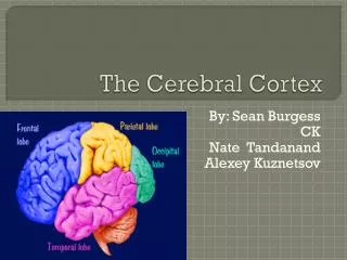
FrontalLobe ParietalLobe PrimaryMotorArea PrimarySensoryArea PremotorArea leg trunk SensoryAssociationArea arm HigherIntellectualFunctions hand VisualAssociation Area face tongue SpeechMotorArea PrimaryVisualArea LanguageComprehension& Formation PrimaryAuditoryArea Cortical functions Learning and memory Memory TemporalLobe OccipitalLobe
Info comes from… • Surgeries • Accidents • Illness • Anatomy (autopsies) • Open brain studies • Animals • Live Imaging
Structures and functions of the brain • Brain imaging techniques • non-invasive techniques include the EEG, CAT, PETT, and the MRI • EEG: Electroencephalography • CAT: Computerized Axial Tomography • PETT: Positron Emission Transaxial Tomography • MRI: Magnetic Resonance Imagery
Stroke Damage fMRI
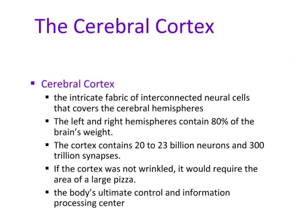
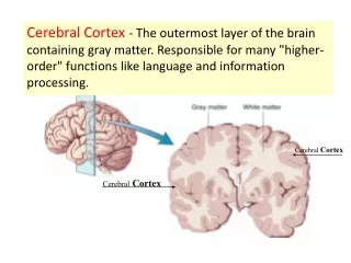
Physiologic anatomy of the cerebral cortex • Cortex is a layer of neurons (2 to 5 mm thick) • Total cerebral cortex contains about 100 billion neurons • Most of the neurons are of three types: • Granular • Fusiform • Pyramidal • Granular neurons with short axons and neurotransmitter types • Output fibers from the cortex (pyramidal and fusiform cells) • Horizontal fibers to adjacent areas and vertical fibers to lower brain areas some all the way down to the spinal cord
Brain • 100 billion neurons. • 10x more glial cells! • Weighs about 1.5 kg, uses 20% of blood flow
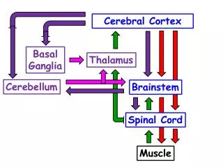
Anatomical and functional relations of the cerebral cortex to the thalamus and other lower centers • Connections to and from the cortex • When the thalamus is damaged along with the cortex, loss of cerebral function is greater • When the thalamic connections are cut, the functions of the corresponding cortical area become almost entirely lost • Thalamo-Cortical system • All sensory information except some sensory pathways of olfaction pass through the thalamus
Anatomical and functional relations of the cerebral cortex to the thalamus and other lower centers • Cortical areas that connect with specific areas of the thalamus
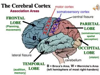
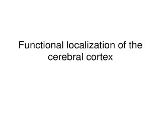
Functions of Specific Cortical Areas • Different cortical areas have separate functions • Electrical stimulation of these areas in patients by Penfield and Rasmussen
Primary sensory and motor areas • The primary motor areas have direct connections with specific muscles for causing discrete muscle movements • Primary sensory areas detect specific sensations • Auditory, visual or somatic • The secondary areas make sense out of the signals in the primary areas • Supplementary and premotor areas function along with the primary motor cortex and basal ganglia to provide “patterns” of motor activity • Interpretation of shape, texture, color, directions of lines and angles, sound tones and sequences in auditory signals
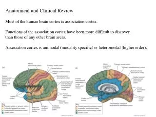
Association Areas • They receive and analyze signals from both motor and sensory cortices as well as from subcortical structures • 1) Parieto-occipitotemporal association areas • 2) Prefrontal association areas • 3) Limbic association area
Parieto-occipitotemporal Association Area • It provides a high level of interpretative meaning for signals from all the surrounding sensory areas • 1) Analysis of the spatial coordinates of the body • 2) Area for language comprehension (Wernicke’s area) • 3) Area for initial processing of visual language (reading): Angular gyrus area • 4) Area for naming objects: Lateral portions of the occipital lobe and posterior temporal lobe
Prefrontal Association Area • It functions in close association with the motor cortex to plan complex patterns and sequences of motor movements • It receives massive subcortical bundle of nerve fibers from parieto-occipitotemporal association area • Prefrontal association area is essential to carrying out “thought” processes in the mind (working memory) • It processes motor and nonmotor information from widespread areas of the brain
Broca’s Area Prefrontal Association Area • Broca’s Area: this area provides neural circuitry for word formation • Located in the posterior lateral prefrontal cortex and partly in the premotor area • Works closely with the Wernicke’s area • Learning one language at a time versus learning two languages at the same time (stored together)
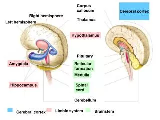
Limbic Association Area • Anterior portion of the temporal lobe, ventral portion of the frontal lobe and in the cyngulate gyrus in the midsurface of each cerebral hemisphere • Behavior, emotions and motivation • Functions of the limbic system
Area for recognition of faces • Prosophenosia: inability to recognise faces • Relationship with the occipital lobe and limbic system
Wernicke’s Area • Comphrensive interpretative function of the posterior superior temporal lobe • Somatic, visual and auditory association areas all meet one another in the posterior part of the superior temporal lobe • Highly developed in the dominant side of the brain • Intelligince area • Damage to Wernicke’s area, one can hear and recognize different words but unable to arrange these words into a coherent thought
Auditory Association Area Wernicke’s Area Wernicke’s Area
Angular Gyrus – Interpretation of Visual Information • Most inferior portion of the posterior parietal lobe, lying immediately behind the Wernicke’s area • Damage to this area, one can still interpret auditory experiences, but stream of visual information into the Wernicke’s area is blocked • Word blindness, Dyslexia • Loss of Wernicke’s area in adults usually leads to a lifetime demented existence
Concenpt of Dominant Hemisphere • Wernicke’s area, angular gyrus as well as functions of the speech and motor control areas are much more highly developed in one cerebral hemisphere than in the other • In 95 % of population the dominant hemisphere is in the left • At birth, size of the Wernicke’s area is about 50% larger than in the right • If the dominant area is damaged during childhood, the opposite side is usually develops dominant characteristics
Concept of Dominant Hemisphere • Dominance in the Broca’s area (the speech area responsible for formation of words by exciting simultaneously the laryngeal, respiratory and mouth muscles • Motor areas for controlling hands are also dominant in 90% of the people (right-handedness) • Role of corpus callosum in communication between the two hemispheres
Role of Language in Functions of the Wernicke’s Area and in Intellectual Functions • A major share of the sensory experience is converted into its language equivalent before being stored in the memory areas or before being processed for other intellectual functions • No storage of visual images of words but as thoughts • Primary and secondary areas of the auiditory areas of the temporal lobe • First introduction to language is by way of hearing
Functions of the parieto-occipitotemporal cortex in the nondominant hemisphere • Damage to dominant hemisphere Wernicke’s area • Many other types of interpretative capabilities of the opposite hemisphere are retained • These areas in the nondominant side may be important for understanding and interpreting music, nonverbal visual experiences, spatial relations of the person with surroundings, body language • Dominant hemisphere, language-based intellectual functions
Higher Intellectual Functions of the Prefrontal Association Areas • It was thought that the prefrontal cortex is the locus of “higher intellect” • Damage to Wernicke’s area and angular gyrus causes more harm to the intellect than that of prefrontal areas • Prefrontal lobotomy and mental changes in patients: • Patients lost their ability to solve complex problems • Unable to string together sequential tasks • Unable to learn to do parallel tasks at the same time
Higher Intellectual Functions of the Prefrontal Association Areas • Their social responses were often inappropriate • They could still talk and comprehend language, but unable to carry long trains of thought, - mood change • The patients could still perform most of the usual patterns of motor function, but often without purpose • Decreased aggresiveness and inappropriate social responses – limbic association area and limbic system • Inability to progress toward goals
Concept of a Working Memory • Performance of higher intellectual functions by prefrontal area • Elaboration of thoughts • Brain’s working memory: • Prognosticate, Plan for the future, Delay action in response to incoming sensory information until the best response is decided; Consider consequences of motor action; Solve complicated mathematical problems; Correlation of info in diagnosing diseases, Control our activities in accord with moral laws
Function of the Brain in Communication • Language input and language output • Sensory and motor aspects of communication
Function of the Brain in Communication • Language input and language output • Sensory and motor aspects of communication
Language • Broca’s area: • articulation of speech. • Damage: slow to speak, comprehension is fine • Wernicke’s area: • language comprehension. • Damage: rapid speech, word salad • To read, hear words: visual, auditory -> Wernicke’s (concept of words) • To speak: Werknicke’s -> arcuate fasciculus -> Broca’s -> motor • Angular gyrus: • Center of integration of auditory, visual, and somatesthetic information.
Function of the Brain in Communication • Wernicke’s aphasia and Global aphasia • Inability to interpret the thought that is expressed (they can still understand spoken or written word) in patients with damage to the Wernicke’s area • Global aphasia: when this damage is widespread and extends to angular gyrus region, lower temporal areas, superior border of the Sylvian fissure
Function of the Brain in Communication • Motor aspects of communication • Process of speech involves two processes: • 1) formation of thoughts in the mind • 2) motor control of vocalization and act of vocalization • Loss of Broca’s area causes motor aphasia • Skilled motor patterns of the vocalizations are lost • Articulation: Facial and laryngeal regions of the motor cortex activate these muscles and the cerebellum, basal ganglia and sensory cortex all help to control sequences and intensities of muscle contractions
Function of the Corpus Callosum and Anterior Commissure • Transfer of thoughts, memories, training and other info • Fibers in the corpus callosum provide abundant bidirectional neural connections • Except the anterior portions of the temporal lobes including amygdala - Anterior Commissure • Role of anterior commissure: emotional responses between the two sides of the brain
Hemispheric Lateralization • Functional differences between left and right hemispheres
Hemispheric Lateralization • Each cerebral hemisphere • Performs certain functions • Not performed by the opposite hemisphere • In most people, left brain (dominant hemisphere) controls: • reading, writing, and math • decision-making • speech and language • Right cerebral hemisphere relates to: • senses (touch, smell, sight, taste, feel) • recognition (faces, voice inflections)
Function of the Corpus Callosum and Anterior Commissure • Cutting CC blocks transfer of info from Wernicke’s area of the dominant side to the motor cortex on the opposite side • Prevents transfer of somatic and visual info from the right side to the Wernicke’s area on the dominant hemisphere • When CC is entirely sectioned, there are two separate conscious portions of the brain • The right side could understand the written word, but not spoken word ...
Thoughts, Consciousness and Memory • Difficulty of studying neural processes involved in thoughts and memory • Removal of cortex does not prevent one from having thoughts, but reduces depth and degree of awareness • Each thought involves simultaneous signals in the cortex, thalamus, limbic system and upper RAS (holistic theory) • Some crude thoughts depend on the lower brain • Pain and stimulation of cortical areas
Thoughts, Consciousness and Memory • Specific stimulated areas of the cerebral cortex determine: • 1) Specific localization of sensations on the body surface and objects in the fields of vision • 2) Feeling the texture of material • 3) Other individual characteristics entering overall awareness • Consciousness: continuing stream of awareness of surroundings or sequential thoughts
Memory (Roles of synaptic facilitation and synaptic inhibition) • Memory storage and changing basic sensitivities of synaptic transmission between neurons • New or facilitated pathways are called memory traces • Memory traces can occur at all levels of the brain • Most memory that we associate with intellectual processes is based on memory traces in cerebral cortex • Positive and negative memory: Sensitization and habituation synaptic transmission
Classification of Memories • 1) Short-term memory • 2) Intermediate long-term memory • 3) long term memory • Working memory – short term memory during intellectual reasoning • Declarative memory • Skill memory is associated with motor activities of the person’s body
Short Term Memory • Short term memory is typified by one’s memory of 7 to 10 numerals in a phone number for a few sec to a few min • As long as the person continues to think about the numbers (facts) • This is caused by continual neural activity resulting from nerve signals that travel around • Presynaptic facilitation or inhibition...
Intermediate Long-Term Memory • It may last for many mins or even weeks • Intermediate long-term memories can result from temporary chemical or physical changes or both in pre- or post-synaptic membrane • Memory based on chemical changes in the pre- and post-synaptic neuronal membrane
Molecular Mehanism of Intermediate Memory • Habituation: results from progressive closure of Ca channels thorugh the terminal membrane • Less Ca ions and less release of sensory terminal transmitter
Molecular Mehanism of Intermediate Memory • Mechanism of facilitation: Release of serotonin • Serotonin acts on its receptors and activation of adenyl cyclase, formation of cAMP • cAMP activates protein kinase – phosphorilation – K channels – blockage of K conductance • Lack of K conductance causes greatly prolonged action potential • Prolonged action potential causes prolonged activation of Ca channels and facilitating synaptic transmission