Download
1 / 14
290 likes | 1.04k Vues
Human Color Perception . By: Lisa Wieczorek Eric King.

E N D
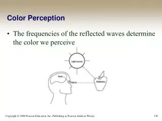
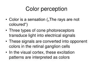
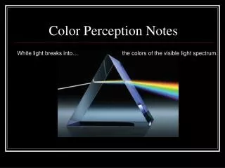
Human Color Perception By: Lisa Wieczorek Eric King • Humans perceive color when light enters the eye and is detected by the photoreceptors, called cones, which are mainly located in the fovea. We have trichromatic color vision which means we have three different kinds of cones that detect a specific wavelength of light, namely short (blue), medium (green), and long (red). How we perceive color also depends on how dim or bright the light is.
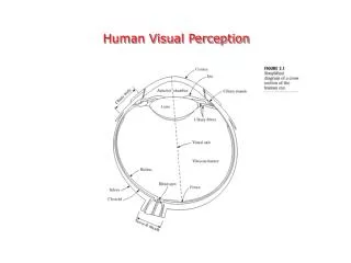
Why can humans see color? • As light enters the eye, it projects an image of the observed object onto the back of the retina. The human eye has the ability to see because of the photoreceptors that convert light into nerve signals. However, as light hits the an area known as the fovea, which has a high density of cones, humans are able to detect color. • Cones: A photoreceptor in the human eye that detects and distinguishes specific wavelengths of light. For humans, there are three types of cones, all of which have different wavelengths of peak sensitivity. The three types of cones are as follows:
Trichromatic Color Vision • Humans have trichromatic color vision which means that they see colors from interactions between three types of cones. • Each of the three cones has a different photosensitive pigment, where each pigment corresponds to a specific wavelength of light. • In 1931, the International Commission on Illumination (CIE) conducted an experiment that had observers look at a white screen through an aperture with a field of view of 2 degrees (the apparent location for the cones in the fovea). The purpose of this experiment was to have the observer manipulate the three primary colored lights on one half of the screen so that when they were mixed together they matched a color that was displayed on the other half of the screen. After obtaining data from multiple “observers” a function was created called the “standard observer”. This function described values of the sensitivity of the red, blue, and green cones to a specific wavelength of light for the average human observer.
Trichromatic Color Vision cont. • The purpose of the “standard observer” is to obtain the coefficients necessary in calculation of the tristimulus values (X,Y,Z). These values represent the combination of red, blue, and green (primary colors) that result in the color being observed. These terms are defined by: • INTEGRALS: • X= Y= Z= • Where represents the source energy multiplied by the reflectance at a given wavelength. • After obtaining X, Y, and Z, the values for the chromaticity are obtained using the following equations: • These values represent the coordinates that are used to determine the object’s observed color in the color space chromaticity diagram. • In using the diagram, only the values of x and y are necessary due to the fact that only two of three coordinates are needed to determine the chromaticity (reccommended by the CIE).
Chromaticity Diagram • This is the diagram used by the CIE to define the chromaticity given chromaticity coordinates x and y. Given a coordinate, one can find the corresponding color in the diagram.
Example of Find Chromaticity: Yellow School Bus • We want to find the color of a school bus when white light is the energy source • First we find the tristimulus values by using the three integrals from before: • X= = 43.7 Y= = 40.3 Z = = 9.8 • Then we calculate the chromaticity by using the other equations for x, y, and z: • = = 0.466 • = = 0.430 • = = 0.104 • These numbers give us our coordinates which are (0.466, 0.430) • Then we find these coordinates on the graph and come up with yellow as the color.
Color Blindness • Color blindness occurs as a result of damage or loss of function of one or more of the cone types. The type of color blindness depends on which of the cone types is compromised. • Dichromacy: One cone type has lost its function. • The most common color blindness as a result of dichromacy is called red-green color blindness which occurs when the middle or the long wavelength cones do not function properly. The result of this is the difficulty in distinguishing between the color red, yellow, and green, but can distinguish between reds and blues. • Another form of dichromatic color blindness (which is very rare) is blue-yellow color blindness due to a defect in the short (blue) cones. In this case, the individual can distinguish between red and green, but cannot see the difference between yellow and blue. • Monochromacy: All three of the cone types are defective • For monochromacy, the individuals cannot see colors, but rather they see shades of black, gray, and white. As a result of this loss of function of the cones, the individuals have a poor visual acuity.
Color Blindness Tests Individuals with blue-yellow color blindness should only see a triangle and not the yellow circle Individuals with red-green color-blindness cannot see the number inscribed the circle
Purkinje Effect • In dim light, we we are most sensitive to wavelengths around 500 nm reflecting on the functions of the rods. In bright light, we are most sensitive to wavelengths around 550 nm reflecting on the functions of the cones. This is the basis for differences in our sensitivities to colors in bright or dim lighting. This is called the Purkinje Effect. Bluish-green object appear brighter in dim light than in bright light and greenish-yellow objects appear brighter in bright light than in dim light. An illustration of the Purkinje Shift. In bright light the eye is more sensitive to longer (red) wavelengths; in dim lighting the eye’s sensitivity shifts slightly to the shorter (blue/violet) wavelengths
Purkinje Effect and Roses • A red rose in daylight makes the red petals appear brighter and the green leaves duller. • In twilight, the red petals appear duller and the green leaves appear brighter.
Bibliography • - The Measurement of Appearance by Richard S. Hunter • - Visual Perception: An introduction by Nicholas J Wade and Michael Swanston • - Human Color Perception by Joseph J. Sheppard, Jr. • http://www.uic.edu/com/eye/LearningAboutVision/EyeFacts/ColorBlindness.shtml • http://www.twodocs.com/color-blind-tests/total-color-blind-test-numbers.htm (red-green test) • http://www.twodocs.com/color-blind-tests/neitz-color-vision-test.htm (blue-yellow) • http://en.wikipedia.org/wiki/Purkinje_effect (rose pic) • http://micro.magnet.fsu.edu/optics/lightandcolor/vision.html • http://photo.net/photo/edscott/vis00010.htm • http://en.wikipedia.org/wiki/Trichromatic_color_vision • http://www.hunterlab.com/pdf/color.pdf • http://www.youtube.com/watch?v=Rab5l5SDm3I
Information Video • http://www.youtube.com/watch?v=Rab5l5SDm3I