Download
1 / 24
370 likes | 1.14k Vues
HEMATOLOGIC (BLOOD) DISEASES. Text Reading Assignment: Chapter 7 - Bleeding Disorders Chapter 8 - Blood Dyscrasias. HEMATOLOGIC (BLOOD) DISEASES. Bleeding Disorders Clotting Factor Disorders Platelet Function Disorders “Blood Dyscrasias” (“Formed Elements”)

E N D
HEMATOLOGIC (BLOOD) DISEASES Text Reading Assignment: Chapter 7 - Bleeding Disorders Chapter 8 - Blood Dyscrasias
HEMATOLOGIC (BLOOD) DISEASES • Bleeding Disorders • Clotting Factor Disorders • Platelet Function Disorders • “Blood Dyscrasias” (“Formed Elements”) • Red Blood Cell (RBC) Disorders • White Blood Cell (WBC) Disorders • Platelet Deficiencies
Bleeding Disorders Platelet Function - Associated Coagulation Factor - Associated Platelet Deficiency Thrombocytopenia / Thrombocytopathia Red Blood Cell (RBC) Disorders Anemia White Blood Cell (WBC) Disorders Leukopenia Leukemia (Lymphoma) HEMATOLOGIC (BLOOD) DISEASES
Oral Bleeding - See Bleeding Protocol Petechiae / Easily Bruised (Ecchymoses) Bleeding After Brushing Spontaneous Gingival Bleeding Prolonged After Extractions Excessive From Minor Trauma Other Bleeding: Epistaxis, Hematemesis, Hemoptysis, Hematuria, Melena Possible Associated Increased Susceptibility to Infection - SeeImmunosuppression Protocol Leukemia HIV Immunosuppression from Chemtherapy for Organ Transplant or Cancer TX Oral Swelling &/or Ulceration Long Term Immunosuppression carries increased Risk for Malignancy (Especially lymphoma and leukemia) CLINICAL FEATURES OF BLOOD DISORDERS Go To Basic Disease
Clinical Bleeding Petechiae Petechiae and Ecchymoses Ecchymoses
Clinical PhotosBleeding(and sometimes Swollen) Gums Leukemia Hemophilia Leukemia Leukemia
Other Clinical Features of Blood Disorders Ulceration Atrophy and Pallor Ulceration
HEMATOLOGIC (BLOOD) DISEASES Health History Correlation Go To Health History
Prothrombin Time (PT) (Extrinsic Pathway) DBL N ~> 25 sec.(N=12-14 sec.) (Activated)Partial Thromboplastin Time (APTT) (Intrinsic Pathway) DBL N ~ > 50 sec.(N=25-35 sec.) International Normalized Ratio(INR) > 3.0 (N=1.0-2.0) Bleeding Time> 10 min. (N~5 min.) Clotting Time > 10 min.(N = < 5 Minutes) Platelet Count as Part of CBC with Differential WBC - < 20 - 40K (N=150-500K/mm3) TESTING FOR BLEEDING DISORDERS INR = PTR ISI = (ProThrombinRatio) ISI = (Patient’s PT/ControlPT)ISI Normal INR = 1.0-2.0 ISI = International Sensitivity Index (for Lab Thromboplastin)
Bleeding Time N ~ 5 min.
Clotting Time N = < 5 Minutes
Primary or Secondary Deficiency of Platelets 10 / Idiopathic (Probably Autoimmune) Thrombocytopenic Purpura 20 / i.e. Leukemia, HIV, Aplastic Anemia Altered Platelet Function as in ASPIRIN (and other NSAID’s) von Willebrands disease Petechiae are Common Finding PLATELET-ASSOCIATED BLEEDING DISORDERSThrombocytopeniaThrombocytopathia (Thrombasthenia)
Hereditary Defects Hemophilia A(VII), B(IX), or C(XI) Other: Parahemophilia (V) and Afibrinogenemia (I) Liver Disease Cirrhosis, Hepatitis (I and II) + (VII, IX, and X) Anti-Coagulant Medication Coumarin (Warfarin) - Vitamin K Antagonist (II, VII, IX, and X) Heparin - Anti-Thrombin / Plasma Thromboplastin GI Malabsorption Problems Fat Soluble Vitamin K Deficiency (Sprue or Biliary Disease) COAGULATION FACTOR-ASSOCIATED BLEEDING DISORDERS
Blood DyscrasiasDiseases of the Formed Elements • Anemia • Leukopenia • Leukemia • (Thrombocytopenia) Test with Complete Blood Count (CBC) with Differential White Cell Count (WBC): RBC Count - Normal = 4.5-5.0 Million RBC’s / 100 mL WBC Count - Normal = 4 - 6 Thousand WBC’s / 100 mL Neutrophils ~ 60 - 65 % Lymphocytes ~ 30 - 35 % Monocytes ~ 4 - 6 % Eosinophils ~ 1 - 2 % Basophils ~ 0 - 1 % Platelets = 150 - 600 Thousand / 100 mL
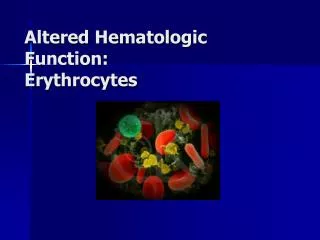
Clinical: Weakness, Fatigue, Pallor Decreased Oxygen Carrying Capacity of Blood Result of: Decreased Number, Size, or HgB Content of RBC’s or of Defective HgB Secondary to: Nutritional / Iron Defeciency RBC loss or destruction (Chronic Bleeding) Failure of RBC formation (Leukemia) Hereditary HgB malformation Oral Features: Pallor Bald Tongue Possible Association with other Disease: Leukemia, Kidney Disease, etc. ANEMIA
Anemia Classification • Size of RBC’s • Microcytic (Small) • Macrocytic (Large) • Normocytic (Normal Size) • Concentration of Hgb • Hypochromic (Less) • Hyperchromic (More) • Normochromic (Normal) • Microcytic / Hypochromic • Chronic Blood Loss, Iron Deficiency, Thalassemia • Macrocytic (Megaloblastic) / Hyperchromic • Vit B12 (Pernicious) or Folic Acid Deficiency • Normocytic / Normochromic • Hemolytic, Aplastic, Myelophthisic, Acute Blood Loss, Chronic Renal Failure
Determination of RBC Indices • RBC count (RBC) - # RBC’s / 100 mL of Blood • (NORMAL = 4.5-5.0 Million / 100 mL) • Hematocrit (Hct) - % of (RB) Cells By Volume • (NORMAL = 36-45%) • Hemoglobin (Hgb) - mg / 100 ml of Blood • (NORMAL = 13-15 mg/dL) • Mean Corpuscular Volume (MCV) • Hct/RBC - Normal = 90 (+or- 10) cubic microliter • Mean Corpuscular Hemoglobin (MCH) • Hgb/RBC - Normal = 30 (+or- 3) picograms • Mean Corpuscular Hgb Concentration (MCHC) • Hgb/Hct - Normal = 33 (+or- 2) %
Aplastic Anemia Failure of Formation of All Blood Cells: RBC’s, WBC’s and Platelets Agranulocytosis Failure of Formation of Neutrophils Cyclic Neutropenia Periodic Suppression of Neutrophil Formation Associated Oral Ulceration and Infection: Candidiasis HSV LEUKOPENIA (Decreased Number of WBC’S)Increased Susceptibility to Infection
Oral Ulceration and Infection Secondary to Leukopenia or Leukemia
Leukemia - Definition Malignancies of WBC’s Originating in BONE MARROW Expression in PERIPHERAL BLOOD Leukemia - Classification Acute Lymphocytic Leukemia (ALL) - Children Chronic Lymphocytic Leukemia (CLL) - Elderly Acute Myelogenous Leukemia (AML) - All Ages Chronic Myelogenous Leukemia (CML) - Adults Clinical Significance Disease and Treatment Make Patients Anemic and More Susceptible to Infection and Bleeding Decreased RBC Formation, IneffectiveLeukocytes (&/orLeukopenia) and Thrombocytopenia Diagnosis by: CBC with Differential and Bone Marrow Biopsy Leukemia - Blood Smear LEUKEMIA Leukemia
Cancer of Lymphocytes Lymph Nodes or Extranodal Soft Tissue (including mouth) - NOT Blood or Bone Marrow Classified by: Hodgkin’s Disease(with REED-STERNBERG CELLS) VS. Non-Hodgkin Lymphoma Type of Lymphocyte: B-cell; T-cell; etc. Maturity of Malignant Cells LYMPHOMA Non-Hodgkin Lymphoma
THE END Test with Complete Blood Count (CBC) with Differential White Cell Count (WBC) + Hgb, Hct, and Red Cell Indices: RBC Count - Normal = 4.5-5.0 Million RBC’s / 100 mL WBC Count - Normal = 4 - 6 Thousand WBC’s / 100 mL Neutrophils ~ 60 - 65 % Lymphocytes ~ 30 - 35 % Monocytes ~ 4 - 6 % Eosinophils ~ 1 - 2 % Basophils ~ 0 - 1 % Platelets = 150 - 600 Thousand / 100 mL