Download
1 / 205
2.22k likes | 2.8k Vues
13 The Brain and Cranial Nerves. Section 1: Functional Anatomy of Brain and Cranial Nerves. Learning Outcomes 13.1 Name the major regions of the brain, and describe their functions.

E N D
13 The Brain and Cranial Nerves
Section 1: Functional Anatomy of Brain and Cranial Nerves Learning Outcomes 13.1 Name the major regions of the brain, and describe their functions. 13.2 Explain how the brain is protected and supported, and how cerebrospinal fluid forms and circulates. 13.3 List the components of the medulla oblongata and pons, and specify the functions of each. 13.4 List the main components of the cerebellum, and specify the functions of each.
Section 1: Functional Anatomy of Brain and Cranial Nerves Learning Outcomes 13.5 List the main components of the midbrain, and specify the functions of each. 13.6 List the main components of the diencephalon, and specify the functions of each. 13.7 Identify the main components of the limbic system, and specify the locations and functions of each. 13.8 Describe the structure and function of the basal nuclei of the cerebrum. 13.9 Identify the major superficial landmarks of the cerebrum, and cite the locations of each.
Section 1: Functional Anatomy of Brain and Cranial Nerves Learning Outcomes 13.10 Identify the locations of the motor, sensory, and association areas of the cerebral cortex, and discuss the functions of each. 13.11 Discuss the significance of the white matter of the cerebral cortex. 13.12CLINICAL MODULE Discuss the origin and significance of the major categories of brain waves seen in an electroencephalogram. 13.13 Identify the cranial nerves by name and number, and cite the functions of each.
Section 1: Functional Anatomy of Brain and Cranial Nerves Brain characteristics Equals ~97% of body’s neural tissue in adults “Typical” brain Weighs 1.4 kg (3 lb) Has volume of 1200 mL (71 in.3) Size varies among individuals Male are ~10% larger than female Owing to differences in overall body size No correlation between size and intelligence Functional normal individuals with smallest (750 mL) and largest (2100 mL) brains
Section 1: Functional Anatomy of Brain and Cranial Nerves Brain development at 4 weeks Neural tube is present Hollow cylinder that is beginning of CNS Has internal passageway (neurocoel) Cephalic portion enlarges into three portions (primary brain vesicles) Prosencephalon (proso, forward + encephalos, brain) “Forebrain” is at tip of neural tube Mesencephalon “Midbrain” is an expansion caudal to prosencephalon Rhombencephalon “Hindbrain” most caudal portion, continuous with spinal cord
Figure 13 Section 1 A lateral view of the brain of an embryo after 4 weeks of development showing the neural tube Mesencephalon (covered by cerebrum) Diencephalon (covered by cerebrum) Rhombencephalon Mesencephalon Cerebrum Spinal cord Prosencephalon Neurocoel A lateral view of the brain of a 5-week-old embryo Prosencephalon Rhombencephalon Pons Metencephalon Myelencephalon Medulla oblongata Diencephalon Cerebellum Spinal cord Telencephalon Spinal cord Brain development in a child, showing the cerebrum covering other portions of the brain
Section 1: Functional Anatomy of Brain and Cranial Nerves Brain development at 5 weeks Primary brain vesicles change position and prosencephalon and rhombencencephalon subdivide to form secondary brain vesicles Prosencephalon Diencephalon (dia, through + encephalos, brain) Becomes major relay and processing center for information to/from cerebrum Telencephalon (telos, end) Becomes cerebrum in adult brain
Section 1: Functional Anatomy of Brain and Cranial Nerves Brain development at 5 weeks (continued) Secondary brain vesicles (continued) Rhombencephalon Metencephalon (meta, after) Adjacent to mesencephalon Forms cerebellum and pons in adult brain Myelencephalon (myelon, spinal cord) Becomes medulla oblongata in adult brain
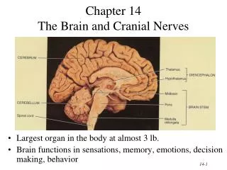
Module 13.1: Major brain regions Major brain regions Cerebrum Divided into pair of large cerebral hemispheres Surfaces covered by superficial layer of gray matter = Cerebral cortex (cortex, rind or bark) Functions Conscious thought Memory storage and processing Regulation of skeletal muscle contractions
Module 13.1: Major brain regions Superficial cerebral structures Fissures Deep grooves that subdivide hemispheres Gyri (singular, gyrus) Folds in cerebral cortex that increase surface area Sulci (singular, sulcus) Shallow depressions in cerebral cortex that separate adjacent gyri
Module 13.1: Major brain regions Cerebellum Partially hidden by cerebral hemispheres Second largest structure of brain Functions Coordination and modulation of motor commands from cerebral cortex
Figure 13.1 1 A diagrammatic view of the brain showing its major regions and their general functions Cerebrum Is divided into a pair of large cerebral hemispheres whose surfaces are covered by a superficial layer of gray matter called the cerebral cortex Fissures Sulci Gyri Diencephalon Is the structural and functional link between the cerebral hemispheres and the rest of the CNS. Thalamus Spinal cord Not visible in this view; the hypothalamus, or floor of the diencephalon Cerebellum Functions in coordination and modulation of motor commands from the cerebral cortex Brain stem Includes three structures Midbrain Medulla oblongata Pons
Module 13.1: Major brain regions Diencephalon Structural and functional link between cerebral hemispheres and rest of CNS Two parts Thalamus Relay and processing centers for sensory information Hypothalamus (hypo-, below) Floor of diencephalon Contains centers involved with Emotions Autonomic function Hormone production
Module 13.1: Major brain regions Brain stem (3 parts) Midbrain Contains nuclei that coordinate visual and auditory reflexes Contains centers that help to maintain consciousness Pons (pons, bridge) Connects cerebellum to brain stem Has tracts and relay centers Contains nuclei that function in somatic and visceral motor control
Module 13.1: Major brain regions Brain stem (3 parts, continued) Medulla oblongata Relays sensory information to other areas of brain stem and thalamus Contains major centers that regulate autonomic function Examples: heart rate, blood pressure Animation: Brain
Figure 13.1 2 Two views of the ventricles, which are filled with cerebrospinal fluid Cerebral hemispheres Cerebral hemispheres Ventricles of the Brain Lateral ventricle Interventricular foramen Third ventricle Aqueduct of midbrain Fourth ventricle Pons Medulla oblongata Central canal Central canal Cerebellum Spinal cord Ventricular system, lateral view Ventricular system, anterior view
Module 13.1: Major brain regions Ventricles of the brain Fluid-filled cavities Filled with cerebrospinal fluid Lined with ependymal cells Formed during development as neurocoel expands within cerebral hemispheres, diencephalon, and metencephalon Connected by narrow canals
Module 13.1: Major brain regions Four ventricles 1. & 2. Lateral ventricles Contained within each cerebral hemisphere Each connected to third ventricle by interventricular foramen Separated medially by septum pellucidum “Roof” partially formed by thick white matter tract connecting hemispheres (corpus callosum) Then narrows to become central canal of spinal cord
Module 13.1: Major brain regions Four ventricles (continued) Third ventricle Contained within diencephalon Connected to fourth ventricle by aqueduct of the midbrain Fourth ventricle Begins in metencephalon and extends into superior portion of medulla oblongata Then narrows to become central canal of spinal cord
Figure 13.1 2 Cerebral hemispheres Ventricles of the Brain Lateral ventricle Interventricular foramen Third ventricle Aqueduct of midbrain Fourth ventricle Pons Medulla oblongata Central canal Spinal cord Ventricular system, lateral view
Figure 13.1 2 Cerebral hemispheres Ventricles of the Brain Lateral ventricle Interventricular foramen Third ventricle Aqueduct of midbrain Fourth ventricle Central canal Cerebellum Ventricular system, anterior view
Figure 13.1 3 Two views of the ventricles, which are filled with cerebrospinal fluid Corpus callosum Lateral ventricles Interventricular foramen Septum pellucidum Third ventricle Inferior tip of lateral ventricle Aqueduct of midbrain Fourth ventricle Cerebellum Central canal
Module 13.1 Review a. Name the major regions of the brain and the distinct structures of each. b. Describe the role of the medulla oblongata. c. Compare the corpus callosum to the septum pellucidum.
Module 13.2: Cranial meninges and cerebrospinal fluid Cranial meninges Dura mater Consists of two layers Separated by slender fluid-filled gap containing fluids and blood vessels Outer (endosteal) layer Fused to cranial bones (no epidural space) Inner (meningeal) layer
Module 13.2: Cranial meninges and cerebrospinal fluid Cranial meninges (continued) Arachnoid mater Consists of Arachnoid membrane Provides smooth covering that does not follow brain’s underlying folds Subarachnoid space lies below Arachnoid trabeculae Connect to pia mater
Module 13.2: Cranial meninges and cerebrospinal fluid Cranial meninges (continued) Pia mater Bound to brain surface by astrocyte processes Extends into every fold and accompanies cerebral blood vessels extending into surface brain structures
Figure 13.2 1 The three layers of the cranial meninges: the dura mater, arachnoid mater, and pia mater Subdural space Cranium (skull) Arachnoid mater Dura mater Arachnoid membrane Dura mater (endosteal layer) Subarachnoid space Dural sinus Arachnoid trabeculae Dura mater (meningeal layer) Pia mater Cerebral cortex Is bound to the surface of the brain by astrocytes
Module 13.2: Cranial meninges and cerebrospinal fluid Dural folds and sinuses Dural folds Dip into cranial cavity and return Provide additional stabilization and support to brain Falx cerebri (falx, sickle shaped) Projects between cerebral hemispheres Inferior attachment to crista galli (anteriorly) and internal occipital crest (posteriorly) Superior and inferior sagittal sinuses lie within
Module 13.2: Cranial meninges and cerebrospinal fluid Dural folds and sinuses (continued) Dural folds (continued) Tentorium cerebelli (tentorium, a tent) Separates cerebrum from cerebellum Falx cerebelli Separates cerebellar hemispheres along midsagittal line Inferior to tentorium cerebelli
Figure 13.2 2 Inferior sagittal sinus Superior sagittal sinus The dural sinuses and dural folds Tentorium cerebelli Falx cerebri Falx cerebelli
Module 13.2: Cranial meninges and cerebrospinal fluid Cerebrospinal fluid (CSF) Completely surrounds and bathes CNS exposed surfaces Materials diffuse between CSF and interstitial fluid of CNS across ependymal walls Total volume = ~150 mL Entire volume replaced in ~8 hours
Module 13.2: Cranial meninges and cerebrospinal fluid Cerebrospinal fluid (continued) Choroid plexus (choroid, vascular coat; plexus, network) Consists of ependymal cells and capillaries Produces CSF ~500 mL/day Found in all ventricles
Module 13.2: Cranial meninges and cerebrospinal fluid Cerebrospinal fluid circulation Created and circulates between ventricles From fourth ventricle, CSF can circulate Down central canal of spinal cord Out single median aperture and lateral apertures into subarachnoid space Down around spinal cord and cauda equina Up around brain Absorbed back into venous circulation through arachnoid granulations within superior sagittal sinus
Figure 13.2 3 The sites of cerebrospinal fluid production, circulation, and absorption into the venous system Superior sagittal sinus Third ventricle Aqueduct of the midbrain Central canal of spinal cord Dura mater Arachnoid Subarachnoid space
Figure 13.2 3 The sites of cerebrospinal fluid production, circulation, and absorption into the venous system Nutrients, O2 Interstitial fluid in thalamus Capillaries Waste products, CO2 Neuron Astrocyte Choroid plexus cells Ependymal cells Removal of waste Cerebrospinal fluid in third ventricle Production of CSF Choroid plexus
Figure 13.2 4 Dura mater Superior sagittal sinus Cranium Arachnoid granulation CSF movement Subdural space Arachnoid membrane Cerebral cortex Pia mater An arachnoid granulation, the site at which cerebrospinal fluid is absorbed into the venous circulation
Module 13.2 Review a. From superficial to deep, name the layers that constitute the cranial meninges. b. What would happen if the normal circulation or reabsorption of CSF became blocked? c. How would decreased diffusion across the arachnoid granulations affect the volume of cerebrospinal fluid in the ventricles?
Module 13.3: Medulla oblongata and pons Medulla oblongata All communication (sensory and motor) between brain and spinal cord passes through Center for coordination of relatively complex autonomic reflexes and control of visceral functions Major anatomical features Olive (prominent bulge and anterolateral surface) Pyramids (contain descending/motor tracts from cerebral cortex) Some fibers cross over to other side of spinal cord = Decussation (decussation, crossing over)
Figure 13.3 1 Structure of the medulla oblongata The anterior surface of the medulla oblongata Pons Pyramids Site of decussation
Module 13.3: Medulla oblongata and pons Medulla oblongata components Autonomic centers (controlling vital functions) Reticular formation Cardiovascular centers Respiratory rhythmicity center Solitary nucleus Relay stations Olivary nucleus Nucleus cuneatus Nucleus gracilis
Figure 13.3 2 Structure of the medulla oblongata Olive Autonomic centers Reticular formation Attachment to membranous roof of fourth ventricle Cardiovascular centers Respiratory rhythmicity center Two views of the structure of the medulla oblongata showing its landmarks and structures Solitary nucleus Relay stations Posterior median sulcus Olivary nucleus Nucleus cuneatus Spinal cord Nucleus gracilis Posterolateral view Anterior view
Module 13.3: Medulla oblongata and pons Pons Links cerebellum with midbrain, diencephalon, cerebrum, medulla oblongata, and spinal cord Contains: Tracts (ascending and descending) Respiratory centers (pneumotaxic and apneustic) Reticular formation Loosely organized mass of gray matter containing centers that regulate vital autonomic functions Extends from medulla oblongata to mesencephalon
Figure 13.3 3 The pons, which links the cerebellum with the midbrain, diencephalon, cerebrum, medulla oblongata, and spinal cord Tracts Respiratory Centers Ascending tracts Descending tracts Pneumotaxic center Apneustic center Transverse fibers Cerebellum Midbrain Fourth ventricle Pons Reticular formation Medulla oblongata Olivary nucleus Spinal cord
Module 13.3 Review a. What is the function of the ascending and descending tracts in the medulla oblongata? b. Name the medulla oblongata parts that relay somatic sensory information to the thalamus. c. Describe the pyramids of the medulla oblongata and the result of decussation.
Module 13.4: Cerebellum Cerebellum Is an automatic processing center that monitors proprioceptive, visual, tactile, balance, and auditory sensations Has two primary functions Adjusting postural muscles Programming and fine-tuning movements controlled at conscious and subconscious levels Ataxia (ataxia, lack of order) Disturbance of muscular coordination from trauma, stroke, or drugs such as alcohol
Module 13.4: Cerebellum Components (posterior view) Anterior and posterior lobes Separated by primary fissure Two hemispheres Separated by vermis (worm) Surface of gray matter (cerebellar cortex) Contains huge, highly branched Purkinje cells that form many sensory and motor synapses Has folds (folia)
Figure 13.4 1 Cerebellum Posterior view Structural features of the cerebellum The posterior, superior surface of the cerebellum Vermis Primary fissure Anterior lobe Posterior lobe Folia Left Hemisphere of Cerebellum Right Hemisphere of Cerebellum