Download
1 / 12
130 likes | 402 Vues
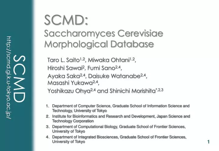
SCMD: Saccharomyces Cerevisiae Morphological Database. Taro L. Saito 1,2 , Miwaka Ohtani 1,2 , Hiroshi Sawai 2 , Fumi Sano 2,4 , Ayaka Saka 2,4 , Daisuke Watanabe 2,4 , Masashi Yukawa 2,4 , Yoshikazu Ohya 2,4 and Shinichi Morishita *,2,3.

E N D
SCMD:Saccharomyces Cerevisiae Morphological Database Taro L. Saito1,2, Miwaka Ohtani1,2, Hiroshi Sawai2, Fumi Sano2,4, Ayaka Saka2,4, Daisuke Watanabe2,4, Masashi Yukawa2,4, Yoshikazu Ohya2,4 and Shinichi Morishita*,2,3 • Department of Computer Science, Graduate School of Information Science and Technology, University of Tokyo • 2. Institute for Bioinformatics and Research and Development, Japan Science and Technology Corporation • 3. Department of Computational Biology, Graduate School of Frontier Sciences, University of Tokyo • 4. Department of Integrated Biosciences, Graduate School of Frontier Sciences, University of Tokyo
What is SCMD ? • SCMD is a collection of micrographs of budding yeast mutants. • Our image processing program, called CalMorph, clips individual cells from micrographs, then analyzes the characteristics of them. • Currently following number of yeast mutants and their micrographs are analyzed. • Mutants analyzed: 4,495 • Micrographs Processed: 84,407 (x 3) • Cells retrieved: 1,643,835
Micrographs • Three type of stains are used to see • Cell wall • FITC-ConA • Nucleus location • DAPI • Actin distribution • Rh-ph • All micrographs can be seen in the viewer provided in SCMD Cell wall Nucleus Actin image processing
stain FITC-ConA DAPI Rh-ph
Categorizing by Bud Size • View of ‘group by bud size’ no bud small medium large yor202w his3
Nucleus Location Categories • We categorized the states of nucleus locations into 7 groups. • State A, A1, B, C are regular status. • State D, E, F seldom occur.
Categorizing by Nucleus Location • View of ‘group by nucleus location’ A A1 B C ybl052c sas3
Categorizing by Actin Localization • View of ‘group by actin localization’ A B iso api E F
Searching Similar Shape Mutants input shape query result