Download
1 / 46
490 likes | 1.09k Vues
Hematopoietic System- 1 Peripheral Blood. Linda F. Cunningham, MD January 2, 2003. Slides. ASCP 1.3 Normal Peripheral Blood Smear. Hematopoietic System Clinical Problems. Anemia Cytosis and Cytopenia Leukocytosis Thrombocytopenia Lymphadenopathy. Hematopoietic System. Blood Plasma

E N D
Hematopoietic System- 1Peripheral Blood Linda F. Cunningham, MD January 2, 2003
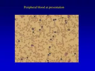
Slides • ASCP 1.3 Normal Peripheral Blood Smear
Hematopoietic SystemClinical Problems • Anemia • Cytosis and Cytopenia • Leukocytosis • Thrombocytopenia • Lymphadenopathy
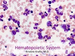
Hematopoietic System • Blood • Plasma • Cells and Proteins • Bone Marrow • Lymphoid Organs • Lymph Nodes, Spleen, and Thymus
Blood • Connective Tissue • Mesodermal Origin • Composition • Cells • Aqueous Plasma Matrix • Proteins • Organic and inorganic salts
Integrated HistologyBlood • Histology of red blood cells (RBCs, erythrocytes), white blood cells (WBCs, leukocytes), and platelets • Composition, biochemistry, and physiology of plasma proteins, inorganic salts, and organic compounds • Fibrinogen for clotting (coagulation)
Slides • ASCP 1.3 Normal Peripheral Blood Smear
Medical HistologyLaboratory Tests Collect blood (venipuncture) in tube without anticoagulant • clotting occurs, using clotting proteins and platelets • aqueous plasma becomes serum • serum is plasma without fibrinogen • serum is sample used to analyze proteins, glucose, etc
Medical HistologyLaboratory Tests Collect blood in tube with an anticoagulant and centrifuge • Two (2) major layers form • Bottom solid cellular layer • Largest layer, red cells, bottom red layer • White cell, “buffy coat” small white/gray layer • Top layer, invisible thin platelet layer • Top aqueous plasma layer
Medical Histology Hematocrit • Volume occupied by red blood cells in the anti-coagulated centrifuged blood • Expressed as % of total volume of blood • Adult male reference range, 39 – 49% • Adult female reference range, 35 – 45%
Blood Cells • Erythrocytes (RBCs) • Leukocytes (WBCs) • Granulocytes • Neutrophils • Eosinophils • Basophils • Monocytes • Lymphocytes • Platelets ASCP 1.08
Slides • ASCP 1.08 Normal Peripheral Blood Smear
Integrated HistologyErythrocytes • RBC size and shape • RBC membrane structure and proteins • RBC cytoplasmic components • RBC maturation and destruction in the circulation • RBC expected lifespan in the circulation
Slides • Histology 2-6.3 a, b, c: Mature red cell peripheral blood smear, disc. 7.5-7.8 micron, eosinophilic cytoplasmic hemoglobin staining; scanning EM; red cell cytoskeleton maintains shape • Robbins 320:rbc membrane • Rodak 29: Schematic red cell membrane
Leukocytes- Granulocytes • Granules Composition and Function • PrimarySpecific Granules • Neutrophils, 60-70 % • Eosinophils, 2-4% • Basophils, 0-1% (Tissue Mast Cells) • Secondary Nonspecific Azurophilic Granules • Granulocyte Circulation and Migration • Tissue Inflammatory Cells
Leukocytes-GranulocytesFunctional Histology • Granulocytes • Neutrophils • Eosinophils • Basophils • Inflammatory response with margination, emigration, tissue action • Phagocytes • Vacuoles and granules • Lysosomes
Slides • Histology 2-6.7 a, b: Neutrophil, multilobed nucleus, pale cytoplasm with few granules; EM granules • 2-6.10 a, b: Eosinophils, bilobed, bright red granules; EM granules with central dense crystalloid • 2-6.11 a, b, c: Basophils and Mast cells, large dark blue granules; Mast cells in tissue; EM histamine released from granules in cytoplasm • Pathology 1-5.3 a, b, c: Tissue neutrophils; margination and emigration • Histology 2-6.5: Neutrophil phagocytosis, ingest extracellular particles; primary granules lysosomal enzymes
MonocytesFunctional Histology • Agranulocytes * (granules by EM) • Size • Cytoplasmic Components • Azurophilic granules are lysosomes • Phagocytes • Circulation and Migration • Macrophages (Histiocytes)
Slides • Histology 2-6.14: Monocyte, large cell, pale vacuolated cytoplasm, irregular nucleus • Harmening 1-32: Monocytes, variation; fine granules, vacuoles, gray-blue cytoplasm; pseudopods • Robbins 377: Tissue monocyte in case of TB Langerhan cell
LymphocytesIntegrated Histology • Size (small, medium, large) • Site of Differentiation and Maturation • Heterogeneous • Major Classes in Circulation • T lymphocytes (T cells) 80% of lymphocytes • B lymphocytes (B cells) 20% of lymphocytes
Slides • Histology 2-6.12 a, b: Small lymphocyte, 6-9 micron, basophilic cytoplasm (ribosomes) , high N/C ratio; EM • Harmening 1-36: Small mature; intermediate size; indented nucleus; large lymphocyte with granules • 1-40: Monocyte and Lymphocyte • 1.39: Lymphocyte and PMN
PlateletsIntegrated Histology • Size • Number • Cytoplasmic Components • Granules • Tubular System • Function • Hemostasis
Medical HistologyLaboratory Evaluation Complete Blood Cell (CBC) count provides information about the absolute and relative numbers of blood cells in a specimen of blood.
Medical HistologyComplete Blood Cell Count (CBC) • Red blood cell count, size, variation in size, and hemoglobin content • White blood cell count and differential into granulocytic cells, lymphocytes, monocytes, and abnormal cells • Platelet count
Slides • Histology 2-6.16 a, b, c, d: Platelets, 1.5-3.5 micron; cell membrane for adhesion, microtubules for aggregation; granules (alpha, dense, lysosomes) • 6.17 a, b: Platelet aggregation after adhering to damaged endothelium
Medical Histology Cell Counts • Cytosis: increased Leukocytosis, neutrophilia, lymphocytosis, erythrocytosis • Cytopenia: decreased Pancytopenia = all Leukopenia = Low WBC count Neutropenia = Low neutrophil count Lymphocytopenia
Leukocytosis • Leukocytosis is a condition in which the leukocyte count is higher than the reference range • Usually affects a specific type of leukocyte- neutrophil, basophil, eosinophil, lymphocyte
Slides • Leukocytosis • Neutrophilic
Leukocytosis • Reactive • Infections, inflammation, necrosis • Others • Malignant • Leukemia (uncontrolled proliferation of leukocytes, causing an accumulation of leukemic cells in bone marrow and blood) • Others
Slides • Reactive = Benign = Self-Limiting • ASCP 1-04 and 1-07 Neutrophilia • ASCP 3-43 Monocytosis • ASCP 3-26, 3-27, 3-33 Lymphocytosis • Essential #138 IM
Slides • ASCP 400X Neutrophils • 1.43 Normal • 3.1 Reactive • 6.1 Chronic Myelocytic Leukemia • ASCP Monocytosis • 3.43 • ASCP Lymphocytosis • 1.12
Patient WBC=100, 000 Neutrophils 86% 86,000 Lymphocytes 4% 4000 Reference Range 4,000 -10,000 50 – 60% 1800 – 7000 30 – 40% 1500 - 4000 Medical Histology Differential WBC Count
Medical Histology Differential WBC Count • Leukocytosis • Neutrophilic leukocytosis (Neutrophilia) • Absolute lymphocyte count within reference range What is the most likely cause?
Slide • Harmening 15-2 Neutrophilia due to a bacterial infection (septicemia)
Patient WBC=100, 000 Neutrophils 7% 7,000 Lymphocytes 93% 9300 Reference Range 4000 -10,000 50 – 60% 1800 – 7000 30 – 40% 1500 - 4000 Medical Histology Differential WBC
Medical Histology Differential WBC • Leukocytosis • Lymphocytosis • Absolute neutrophil count within reference range What is the most likely cause?
Slide • ASCP 3-40 Lymphocytosis
Medical HistologyPeripheral Blood • Review histology of peripheral blood and give clinical examples of specific leukocytosis • Give examples of a reactive leukocytosis, acute leukemia, and chronic leukemia • Histology is important for the recognition of maturation of leukemic cells
Medical Histology Thrombocytopenia • Decreased platelets • Interrupt blood coagulation and prevent hemostasis • Bleeding complications • The degree of thrombocytopenia determines the risk for bleeding due to minor trauma, risk of spontaneous bleeding, and severe bleeding
Medical Histology Thrombocytopenia • Decreased platelets • Interrupt blood coagulation and prevent hemostasis • Bleeding complications • The degree of thrombocytopenia determines the risk for bleeding due to minor trauma, risk of spontaneous bleeding, and severe bleeding
Medical Histology Anemia Decrease in the total number of circulating erythrocytes and/or a decrease in the quality or quantity of hemoglobin. Number and severity of the symptoms depend upon the body’s ability to compensate for the reduced oxygen carrying capacity of the blood.
Medical Histology Anemia • Decreased Production ( Look in the Bone Marrow) • Hemoglobin synthesis • DNA synthesis and nuclear maturation • Stem cell defects • Bone marrow infiltration
Medical Histology Anemia • Acute Blood Loss (Hemorrhage) • Increased Destruction (Hemolysis) • Membrane abnormality • Cytoplasmic enzyme abnormality • Hemoglobin abnormality • RBC environment (Extracellular)
Medical HistologyAnemia-Morphology • Erythrocyte size = -cytic • Normocytic, macrocytic, microcytic • Hemoglobin content= -chromic • Normochromic, hypochromic
Slide • Normal red blood cells = normocytic for comparison
Slides • Rodak # 86 Iron Deficiency Microcytic • ASCP 4-16 Iron Deficiency Microcytic • ASCP 4- 20 Macrocytic with Lymphocyte • ASCP 4.12 Anisocytosis