Download
1 / 13
210 likes | 674 Vues
Abdominal Ultrasound in Liver Fibrosis. Laurentius A. Lesmana Department of Internal Medicine, University of Indonesia Digestive Disease Centre, Medistra Hospital, Jakarta. US evaluation of the liver.

E N D
Abdominal Ultrasound in Liver Fibrosis Laurentius A. Lesmana Department of Internal Medicine, University of Indonesia Digestive Disease Centre, Medistra Hospital, Jakarta
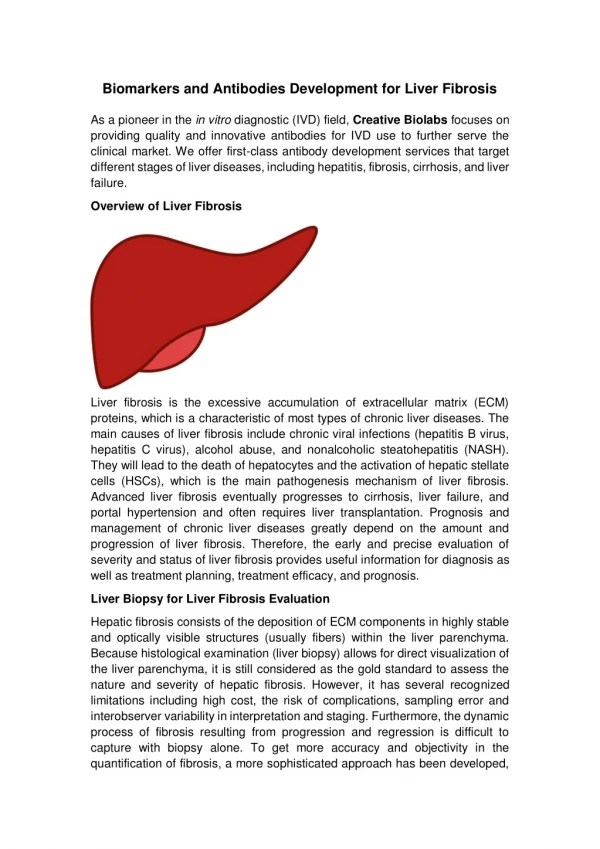
USevaluation of theliver • Adequate scanning technique demands that each patient be examined with the following assessments: • The size of the liver in the longitudinal plane • The attenuation of the liver parenchyma • Liver echotexture • Hepatic vascular structures • Intrahepatic biliary ducts structures
Liver Biopsy – Gold Standard • Obtained by either a percutaneousor US-guided fine-needle aspiration (FNA). • METAVIR Scores: • Fibrosis stage: F1-F4 (cirrhosis) • Inflammatory activity: A1-A3 • Biopsy is NOT NECESSARYif the clinical, laboratory, and radiologic data strongly suggest the presence of cirrhosis.
Liver Fibrosis Staging Diagnostic performance of three US parameters to detect early liver cirrhosis. ( 324 patients were evaluated by both US and liver biopsy ) Shen L, et al. World J Gastroenterol. 2006;12:1292-5.
Accuracy of routine US in staging of liver fibrosis in chronic hepatitis • A retrospective evaluation of ultrasound images in 156 patients with Chronic viral hepatitis. • Three US features were assessed : surface nodularity, liver edge and parenchymal echotexture. • Scores of 0 to 3 ( 0=normal, 1=mild, 2=moderate and 3=severe. . Choong CC, et al. J Clin Imaging Sci. 2012;2:58
US images and scoring system Choong CC, et al. J Clin Imaging Sci. 2012;2:58.
Liver Fibrosis Staging Diagnostic performance of US parameters to detect significant fibrosis (>F2). Choong CC, et al. J Clin Imaging Sci. 2012;2:58.
Liver Fibrosis Staging Diagnostic performance of US parameters to detect severe fibrosis (>F3). Choong CC, et al. J Clin Imaging Sci. 2012;2:58.
Liver Fibrosis Staging Diagnostic performance of US parameters to detect cirrhosis (F4). Choong CC, et al. J Clin Imaging Sci. 2012;2:58.
Conclusion • Conventional US cannot differentiate accurately the different liver fibrosis stage. • Newer ultrasound technique elastography • There are 4 US elastography techniques: • transient elastography (TE), • real time elastography (RTE) • acoustic radiation force impulse imaging (ARFI) • shear wave elastography (SWE)
Recommendations • Conventional ultrasound can not be used as an accurate predictor of early and significant fibrosis in chronic viral hepatitis except for early cirrhosis. • Conventional US is still useful in detecting cirrhosis.