Download
1 / 130
1.4k likes | 1.89k Vues
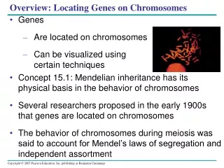
Organization of genes on chromosomes. Mapping genes on chromosome: Genetic mapping (linkage and recombination analyses). Cytogenetics method. Restriction mapping, deletion studies, and other molecular approaches such as chromosome walking. Sequencing. Deduction of gene structure.

E N D
Organization of genes on chromosomes • Mapping genes on chromosome: • Genetic mapping (linkage and recombination analyses). • Cytogenetics method. • Restriction mapping, deletion studies, and other molecular approaches such as chromosomewalking. • Sequencing. • Deduction of gene structure.
Restriction Fragment Length Polymorphism (RFLP) RFLP and VNTR (variable lengths of tandem repeats) are useful in DNA fingerprinting.
Deduction of gene structure • By hybridization of mRNA and chromosomal DNA. • By restriction mapping. • By DNA sequence.
Organization of interrupted genes may be deduced by restriction mapping of cDNA and genomic DNA Small exons or introns may be missed by such an analysis. Resolution at the sequence level is necessary to identify all segments of the gene.
Figure 4.12: Exons are identified by flanking sequences and ORFs.
Introns of nuclear genes generally have termination codons in all reading frames, and have no coding function.
Organization of interrupted genes may be conserved: all globin genes have the following interrupted structures.
Another example with the DHFR gene. Exon sequences are conserved but introns vary
Two genes may share the same sequence by reading the DNA in different frames
Alternative splicing of mRNA can produce more than one protein
Genes can be identified and isolated by many approaches • By classical genetic approach. • By reverse genetic approach. • DNA cloning and/or cDNA approach. • By conservation of exons (eg., zoo blotting). • By analysis of DNA sequence (ORF flanked by splicing junction). • By exon trapping. • By chromosome walking (especially for large-sized gene).
How would you know that a segment of DNA is part of a gene? • Cross-hybridization with • the genomes of other • species (Zoo blot). • 2. Contains open reading • frames (ORF).
Characterization of dystrophin gene by zoo blotting, cDNA identification and chromosomal walking.
How did interrupted genes evolve? Introns early or introns late? 1. The equation of at least some exons with protein domains, and the appearance of related exons in different proteins, indicates that the duplication and juxtaposition of exons has played an important role in evolution. 2. Most protein-coding genes probably originated in an interrupted form, but interrupted genes that code for RNA have originally been uninterrupted.
Every exon of immuoglobulin gene corresponds exactly with a know functional domain of the protein
Chromosomes, nucleosomes and controlling chromatin sturcture • How is DNA packed in the chromosomes. • Unusual chromosome structures. • Nucleosomes. • Controlling chromatin structure. • Gene function and chromatin structure. • Epigenetics (read Chapter 31 of Genes IX) .
DNA Topology • Topology is a branch of mathematics that studies the properties of an object that do not change under continuous deformations. For circular DNA molecules, a topological property is one that is unaffected by deformations of the DNA strands as long as no breaks are introduced. DNA topology is the study of the spatial relationship of DNA. • Topology of DNA • Intramolecular properties – relationship between the two strands of the duplex. Only CCC DNA or linear duplex with at least two anchors are of topological concerns. • Intermolecular properties – relationship between two molecules, eg., catenanes.
Supercoiling of DNA can only occur in closed-circular DNA or linear DNA where the ends are fixed. Underwinding produces negative supercoils, whereas overwinding produces positive supercoils.
Negative and positive supercoils . Topoisomerases catalyze changes in the linking number of DNA.
Topology of cccDNA is defined by: Lk = Tw + Wr, where Lk is the linking number, Tw is twist and Wr is writhe.
Intertwining of the two strands • Nodes = ss crossing on 2D projection. Right-handed crossing = +1/2 Left-handed crossing = -1/2 Lk = number of times one strand winds around the other on 2D projection. One linking number = 2 nodes.
DNA Compaction Requires Solenoidal Supercoiling, not plectonemic supercoiling.
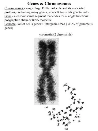
Chromosome, chromatin, chromatid • Chromatin: the complex of DNA and protein in the nucleus of the interphase cell. Heterochromatin refers to regions of the genome that are permanently in a highly condensed condition, while euchromatin refers to the rest of the genome. • Chromosome: consists of one DNA molecule and proteins. Visible as morphological entity only during mitosis. • Chromatids: copies of a chromosome produced by replication.
Packaging of DNA Packing ratio: the length of the DNA divided by the length of the unit that contains it.
HU and H1 proteins may be • involved in condensing DNA. • There are about 400 domains • of independent supercoiling, • each consists of ~10-40 kb. • The average density of • supercoiling is ~1 turn/100 bp. • 4. Treatment with reagents that • act on RNA or protein may • unfold the nucleoid. • Question: How is #2-3 determined?
Loops, domains and scaffolds in eukaryotic DNA • Genome, when isolated carefully, can be visualized as 10 nm fiber, consisting of DNA and protein. Supercoiling measured by EtBr indicates about 1 negative supercoils per 200 bp. • Loops can be seen directly when the majority of histones are removed (see next Fig.). Threads of DNA emanate from the scaffold as loops of average length 10-30 mm (30-90 kb).