Download
1 / 40
420 likes | 454 Vues
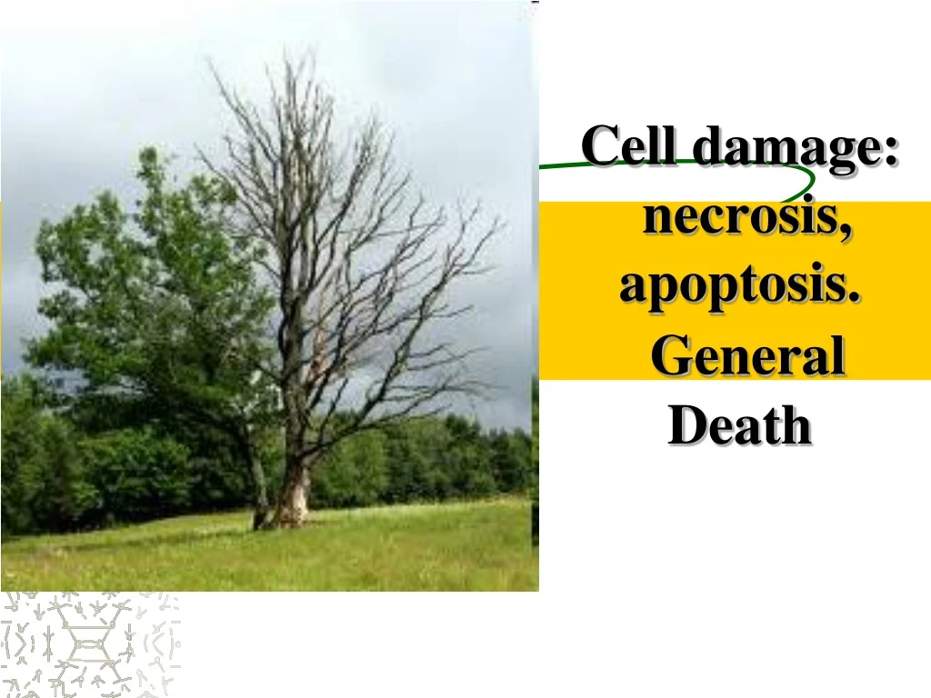
Cell damage: necrosis, apoptosis. General D eath. Necrosis (from the Greek. Nekros - dead) . It’s death of cells and tissues in living organism. Necrosis may be in one cell, a group of cells, part of the body or organ. Stages. 1 - Preneс rosis or paranecrosis 2 - N ecrobiosis

E N D
Necrosis (from the Greek. Nekros - dead). It’sdeath of cells and tissues in living organism. Necrosis may be in one cell, a group of cells, part of the body or organ.
Stages 1 - Preneсrosisor paranecrosis 2 - Necrobiosis 3 - Actually necrosis 4 - Autolysis
Preneсrosisor paranecrosis- changes similar to necrotic, but reverse
Necrobiosis- profound degenerative changes, in which the prevailing catabolic over anabolic changes
Actually necrosis –cell death when the time of death can not be established
Autolysis- decomposition of dead substrate under the influence of hydrolytic enzymes
1.Kariopiknosis - wrinkling nucleus (Я), caused by condensation of chromatin Microscopic signs of necrosis. Changes in the nucleus
2.Karyorhexis – breakdown of nucleus Microscopic signs of necrosis. Changes in the nucleus
nuclei lymph follicleappear as small fragments Microscopic signs of necrosis. Changes in the nucleus– kariorhexis inlymph follicle
3. Kariolysis - total splitting of the nucleus by hydrolytic enzymes Microscopic signs of necrosis. Changes in the nucleus
1.Plasma-coagulation:protein denaturation and coagulation 2. Plasmorhexis -cytoplasm decomposition 3. Plasmolysis - hydrolytic fusion of cytoplasm The process ends with a complete dissolution of cells Microscopic signs of necrosis. Cytoplasm changes
In the interstitial substance of the intercellular space depolymerization with glucosamineglycane and saturation with blood plasma proteins develops. As a result interstitial substance becomes swollen and fuses. The collagen and elastic fibers are fusedtoo. Reticular fibers are preserved longer than the other structures. Then they dissociate to lumps. Microscopic signs of necrosis. Changes intercellular substance
Classification of necrosis I.According to the cause: • 1) traumatic (caused by chemical or physical factors); • 2) toxic (toxins of bacteria, chemicals); • 3) trophoneurotic (in disturbances of nervous trophic)- bed-sore • 4) allergic (develops in the sensitized organism as hypersensitivity reaction of immediate type); • 5) vascular (infarction).
Classification of necrosis II. According to the clinic and morphological forms: • 1) coagulative (dry) necrosis; • 2) colliquative (liquefactive) necrosis; • 3) gangrene (originate from Greek <<gangraina>> fire) necrosis of tissue adjacent to the outer environment: a) dry b) wet • 4) sequestration; • 5) infarction.
Classification of necrosis III. According to mechanism of its development it may be direct and indirect. • Direct necrosis arise immediate action of causal factor (toxic and traumatic); • Indirect (mediate) necrosis may be vascular and trophoneurotic.
This is the most common type of necrosis which is caused by ischemia, and less often by bacterial and chemical agents. The organs commonly affected are the heart, kidneys and spleen. Coagulative necrosis
In gross examination the foci of coagulative necrosis in the early stage are pale, firm and slightly swollen. Microscopic change probably results from denaturation of structural and enzymatic proteins Coagulative necrosis
Colliquative necrosis due to ischemic injury and bacterial infections. Because of hydration and colliquation of tissue by the action of powerful hydrolytic enzymes. The common examples are brain infarct and abscess cavity. Colliquative necrosis
In gross examination the affected area is soft and swollen. Late a cyst wall is formed. Microscopically, the cystic space contains necrotic cell detritus. Macrophages filled with phagocytosed material. Colliquative necrosis
Infarction is vascular necrosis. The causes of infarction are prolonged vascular spasm, thrombosis or embolism . Infarction
Sequestration is an area of dead tissue which does not experience autolization and is freely located in the living tissue. It is characteristic for osteomyelitis - purulent inflammation of the bone marrow. Sequestration
Wet gangrene. Occurs under the action of putrefactive microorganisms. Tissue to swell, become swollen, emit a fetid smell, demarcation zone is not defined. Wet gangrene occur in the lungs, intestines and uterus. Gangrene
Dry gangrene begins in the distal part of a limb due to ischemia. The typical example is the dry gangrene in the toes and feet of an old patient due to arteriosclerosis. Gangrene
Is a kind of gangrene, death of the tissue under the influence of pressure (sacral area, spinous processes, great trochanter). It is trophoneurotic necrosis in severily ill patients. Bed-sore
Outcome of necrosis Favorable: • organization, replacement by connective tissue with formation of a scar or a capsule; • petrifaction; • ossification, formation of bone; • aseptic autolysis. Unfavorable: saprogenic fusion of necrotic tissue followed by sepsis.
APOPTOSIS • Apoptosis - genetically programmed necrosis unwanted or defective cells in living organism and is aimed at the destruction of cells during embryogenesis and physiological involution: death epithelium of the skin, red and white blood cells. • Biological meaning of apoptosis is the elimination of damaged cells. • Between 50 and 70 billion cells die each day due to apoptosis in the average human adult.
These changes include : - blebbing, cell shrinkage; - nuclear fragmentation; - chromatin condensation and chromosomal DNA fragmentation; - formation of apoptotic bodies; - phagocytosis of apoptotic bodies by macrophages or parenchymal cells. Morphogenesis of apoptosis
In contrast to necrosis, which is a form of traumatic cell death that results from acute cellular injury, apoptosis, in general, confers advantages during an organism's life cycle.
Death • Depending on the cause distinguish natural (physiological) death from old age and wear on the body, violent death from injury or other negative effects on the body ending in death, and from diseases (inviolent)
Stages of dying • Agony • Clinical (somatic ) death • Biological (molecular) death
Agony Terminal condition characterized: • Violation of the central nervous system (sopor or coma) • Low blood pressure • Centralization circulatory • Breathing disorders It was during the agony of the body loses 60 ... 80 grams of weight (due to complete combustion and depletion of cellular ATP reserves), which in some scientific-sounding articles called weighing souls who left after the agonyof the body.
Clinical death • Clinical death is characterized by respiratory and cardiac arrest during 5-6 minutes while live brain cells. Clinical death is reversible process of dying. Reversibility depends on the degree of hypoxic changes in the brain.
Biological death • Biological death is the permanent (irreversible) cessation of all biological functions that sustain a living organism.
Relative Relative signs of death: Byeloglazov’s sign (cat eye) While squeezing the eyeball from a deceased pupil becomes oval. Signs of biological death
early: postmortem lividity (PML) changes in the muscles (Cadaveric Rigidity) cooling of the body desiccation of the skin late: preserving of thebody- -saponification оr (Adipocereformation), -mummification Which destroy the body - -putrefaction Reliablesigns of death
Postmortem Lividity (PML) • After termination of cardiac activity and loss of tone of the vascular wall is a passive movement of blood through the vessels by gravity and concentration it in below the body parts.
Mummification • Mummification – means a modification of process of putrefaction in which there is a dehydration or dessication of all body after death.
And yet life is beautiful Thank you for your attention