Download
1 / 94
1.07k likes | 1.44k Vues
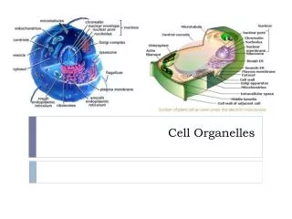
Cell Organelles. Class Presentation on Topic 2 4A– IB Biology HL . * RiBOSOMES *. Definition: Small spherical structures, consisting of two subunits. Key: ABC – from the HLB textbook ABC – from the AP Bio textbook

E N D
Cell Organelles Class Presentation on Topic 2 4A– IB Biology HL
*RiBOSOMES* Definition: Small spherical structures, consisting of two subunits. Key: ABC – from the HLB textbook ABC – from the AP Bio textbook *some sentences may contain the pg. #’s from where the info is derived from. Angel Alabe/Abbey Currence
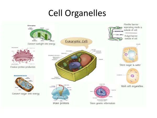
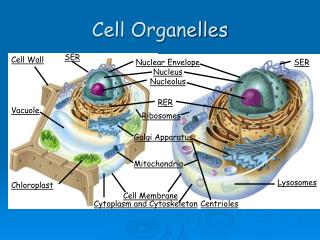
RIBOSOMESStructure & Function • -Carry out protein synthesis. (More protein synthesis, more ribosomes found) • -One large subunit on top with one small subunit attached to bottom • -Occur in all prokaryotic cells and function as sites of protein synthesis • -They occur in very large numbers in cells with high protein production, and when numerous, impart a granular appearance to an electron micrograph of the prokaryotic cell. Larger subunit Small subunit
RIBOSOMESStructure & Function (contd.) • -Ribosomes are complex structures within the plasma membrane, but they have no exterior membrane. • -The part of the cell where proteins are made. Made of rRNA and have two subunits (513) • -Rough ER has ribosomes on the exterior of the channels (22) • -Decode strands of mRNA to produce polypeptides in the space between the two subunits. (204)
RIBOSOMESStructure & Function (contd.) • -Between subunits: binding sites for mRNA and tRNA (site A, P, and E) (204) • A: holds rRNA carrying the next amino acid to be added to the polypeptide chain • P: holds tRNA carrying the growing polypeptide chain • E: site from which tRNA that has lost its amino acid is discharged
RIBOSOMESContents • -Composed of a type of RNA and protein. (22) • -Subunits composed of ribosomal RNA (rRNA) molecules and many distinct proteins. (203) • -About 2/3 ribosomes mass is rRNA. (203)
RIBOSOMESCharacteristics • -One large subunit on top w/ smaller one on bottom • -Molecules of ribosomes are constructed in the nucleolus of eukaryotic cells and exit the nucleus through the membrane pores. (204)
RIBOSOMESProteins Produced: Free vs. Attached • -Both structurally identical • -Free: suspended in cytosol (cytoplasm) • -Bound: attached to outside of endoplasmic reticulum or nuclear envelope • Generally make proteins that are destined for insertion into membranes, for packaging within certain organelles (such as lysosomes), or for export from the cell (secretion)
RIBOSOMESProkaryotic vs. Eukaryotic Cell’s Ribosomes • Euk: Larger and denser than Prok. (22) • Subunits equal 80S (22) • Animal -Small structures, free in the cytoplasm or associated with the endoplasmic reticulum (ER) • Plant –Small (20nm) stuctures which manufacture proteins. May be free in the cytoplasm or associated with the surface of the endoplasmic reticulum • Prok: Smaller and less dense than Euk (22) • Subunits equal 70S • Difference in molecular makeup (204)
RIBOSOMES70S Ribosomes • -Prok. Subunits together equal 70S (22) • -Euk. Subunits together equal 80S • (S= Svedberg units, which indicate the relative rate of sedimentation during high-speed centrifugation) (22)
RIBOSOMESTranslation & Channeling of Proteins *addressing codons* -codons carry the genetic code from DNA to the ribosomes via mRNA. -there are 64 possible codons. -three codons have no complementary tRNAanticodon ( referred to as stop codons) -there is a start codon (AUG) that signals the beginning of a polypeptide chain. -start codon also encodes the amino acid methionine. Translation – The process of producing a protein once mRNA has been produced from the DNA template. (RNA protein) • The process is referred to as translation because it changes the language of DNA to the language of protein • The centre of this process is the ribosome. • Process involves several phases: • Initiation • Elongation • Translocation • Termination co·don noun /ˈkōˌdän/ A sequence of three nucleotides which together form a unit of genetic code in a DNA or RNA molecule
RIBOSOMESDifferences Between Free & rERRibosomes rER – rough endoplasmic reticulum • Free Ribosomesvs. • Move freely in the cytoplasm • Proteins produced are primarily used within the cell (206) • rERRibosomes • Attached to the exterior channels of rER • Involved in protein synthesis • Proteins produced are primarily secreted from the cell or used in lysosomes (206)
RIBOSOMESTranslation & Channeling of Proteins (contd.) • INITIATION PHASE • The start codon (AUG) is on the 5’ end on all mRNAs. • Each codon, other than the three stop codons, attaches to a particular tRNA. The tRNA has a 5’end and a 3’ end like all other nucleic acid strands. • The 3’ end of tRNA is free and has the base sequence of CCA which is the site of amino acid attachment. • Hydrogen bonds form in four areas because there are complementary bases in the single stranded tRNA, which causes the tRNA to fold and tane-on a three dimensional structure. • If the molecule is flattened, it has a two dimensional appearance of a clover leaf. • One of the loops of the clover leaf contains an exposed anticodon which is unique to each type of tRNA. It’s also the anitcodon that pair with a specific codon of mRNA
RIBOSOMESTranslation & Channeling of Proteins (contd.) • INITIATION PHASE • Each of the 20 amino acids binds to the appropriate tRNA due to the action of particular enzyme • Because there are 20 amino acids, there are 20 enzymes • The active site of each enzyme allows a fit only between a specific amino acid and the specific tRNA. • The actual attachment of the amino acid and the tRNA requires energy that is supplied by the ATP. • At this point, the structure , structure is called an activated amino acid, and the tRNA may now deliver the amino acid to a ribosome to produce a polypeptide chain.
RIBOSOMESTranslation & Channeling of Proteins (contd.) • INITIATION PHASE • The initiation phase are the first steps of the Translation process: • When an activated amino acid – methionine attached to a tRNA with the anitcodon UAC – combines with an mRNA strand and a small ribosome unit. • The small subunit moves down the mRNA untill it contact the start codon (AUG), this contact starts the translation process. • Hydrogen bonds form between the initiator tRNA and the start codon • Next, a large ribosomal subunit combines with these parts to form the translation initiation process complex. • Joining the initiation complex are proteins called initiation factors that require energy from guanosinetriphosphate (GTP) for attachment. GTP is an energy-rich compound very similar to ATP
RIBOSOMESTranslation & Channeling of Proteins (contd.) • ELONGATION PHASE Once initiation is complete, elongation occurs • Phase involves tRNAs bringing amino acids to the mRNA – ribosomal complex in the order of specified by the codons of the mRNA. • Proteins called elongation factors assist in the binding the tRNAs to the exposed mRNA codons at the A site. • The initiator tRNA then moves to the P site. The ribosomescatalyse the formation of peptide bonds between adjacent amino acids brought to the polypeptide assembling area.
RIBOSOMESTranslation & Channeling of Proteins (contd.) • TRANSLOCATION PHASE This phase happens during the elongation phase • Involves the movement of the tRNAs from one site of the mRNA to another. • First, a tRNA binds with the A site & its amino acid is then added to the growing polypeptide chain by a peptide bond. • This causes the polypeptide chain to be attached to the tRNA at the A site. • The tRNA then moves to the P site & transfers its polypeptide chain to the new tRNA that moves into the now exposed A site. • Empty tRNA is transferred to the E where it’s released. This process occurs in the 5’ to 3’ direction. • Therefore, the ribosomal complex is moving along the mRNA toward the 3’ end. • Remember that the start codon was near the 5’ end of the mRNA.
RIBOSOMESTranslation & Channeling of Proteins (contd.) TRANSLOCATION PHASE
RIBOSOMESTranslation & Channeling of Proteins (contd.) • TERMINATION PHASE Termination phase completes the translation process • Begins when one of the three stop codons appears at the open of A site • A protein called a release factor then fills the A site (protein factor does not carry an amino acid). • It catalyses the hydrolysis of the bond linking the tRNA in the P site with the polypeptide chain. • This frees the polypeptide, releasing it from the ribosome. • Ribosome then separates frmom the mRNA & splits into its 2 subunits. • Proteins synthesized in this manner have several different destinations. • If they are produced by free ribosomes, proteins are primarily used within the cell. • If produced by ribosomes bound to the ER, they’re primarily secreted from the cell or used in lysosomes.
RIBOSOMESTranslation & Channeling of Proteins (contd.) TERMINATION PHASE
Rough Endoplasmic Reticulum Structure • A membranous system of interconnected tubules and flattened sacs called cisternae. • Rough Endoplasmic Reticulum – studded on its outer surface with ribosomes.
Rough Endoplasmic Reticulum Function • It transports materials throughout the internal region to cell. • Membrane factory for cells; it grows in place by adding membrane proteins and phospholipids to its membrane • It is involved in protein development and transport.
Rough Endoplasmic Reticulum Content • Most secretory proteins in the organelle are glycoprotein (proteins that have carbohydrates covalently bonded to them). The carbohydrates are then attached to the proteins the rough endoplasmic reticulum by specialized molecules built into its membrane.
Rough Endoplasmic Reticulum Rough Endoplasmic Reticulum
Cell Membrane • Definition: The organelle that “functions as a selective barrier or boundary for every cell, and regulates the sufficient passage of oxygen, nutrients, and wastes to service the entire cell.” • Also known as: • Membrane • Plasma membrane • Cell surface membrane • Organelles have membranes, too, that are similar in structure
Cell Membrane Micrograph image of part of a cell: I = Membrane (of the nuclear envelope) II = Mitochondrion
Cell MembranePhospholipid Structure • Structure Overview: There is a bilayer (double layer) in which lipids (fats) stay on the outer part, and proteins pass through the middle among the lipid tails • Phospholipids: Made up of glycerol (3 carbon compound) • 2 of glycerol carbons are attached to fatty acids • 3rd glycerol carbon attached to highly polar organic alcohol (related to phosphate)
Cell MembranePhospholipid Structure • Has a hydrophilic (polar, water soluble) head • Has two hydrophobic (nonpolar, water insoluble) tails • The tails have a weak bond, causing the cell membrane to be very flexible • The heads are connected by hydrogen bonds, and they maintain the structure of the membrane
Cell MembranePhospholipid Structure Cholesterol: • Found in animal cells ONLY • Located in the tails of phospholipids • Determines the membrane fluidity • Membranes MUST be fluid in order to function properly • Consistency is similar to olive oil • Temperature change affects fluidity • Instead of cholesterol, plant cells use unsaturated and saturated fatty acids
Cell MembraneProteins • Many different varieties – Also known as amino acids • Two most prominent types are: • Integral proteins • Peripheral proteins
Cell MembraneProteins Integral: • Hydrophilic regions – polar amino acids (outer regions) • Hydrophobic regions – nonpolar amino acids (central regions) • Regulate the entrance and exit of molecules through the cell membrane I = Integral Protein
Cell MembraneProteins Peripheral: • Stays on the surface of cell membrane • Attached to integral proteins • Also attach to glycoproteins (another type )
Cell Membrane I = Glycoprotein II = Integral protein III = Hydrophilic phosphate head
Cell MembraneProtein Functions 6 basic functions of proteins: • Enzymatic action: relates to metabolic reactions • Cell adhesion: when proteins connect (junctions) • Cell-to-cell communication: for identification • Hormone binding sites • Passive transport channels • Active transport pumps
Cell MembraneTransport Two types of transport are: • Passive transport: movement due to redistribution for different concentrations, no energy expended • Diffusion • Osmosis • Active transport: movement against concentration gradient, energy is expended (in the form of ATP)
Cell MembranePassive Transport Diffusion: movement of non-water particles from a concentration that is high to one that is low • Movement between protein channels (phospholipid molecules) • Facilitated diffusion: carrier proteins combine with substances to help with movement – protein shapechanges but no energy is required still
Cell MembranePassive Transport • Exam Question Example: Channel Proteins • The diagram below shows a channel protein in a membrane. Which parts of the surface of the protein would be composed of polar amino acids? • A. I and II only • B. II and III only • C. III and IV only • D. I and IV only (Answer is A)
Cell MembranePassive Transport Osmosis: • Happens along the concentration gradient • Involves ONLY water molecules • Happens with partially permeable membranes • Movement caused by concentrationdifference inside and outside the cell membrane • Goal of passive transport is to reach equilibrium on both sides
Cell MembranePassive Transport Osmosis: • Hyperosmotic – higher concentration • Hypo-osmotic – lower concentration • Water moves from hypo-osmotic → hyperosmotic • Iso-osmotic – equilibrium, no movement
Cell MembranePassive Transport Osmosis: influencing factors of substances that are being transported are… • Size – small = easy movement large = difficult movement • Charge – nonpolar = easy movement polar = difficult movement
Cell MembranePassive Transport (Partially permeable) Uses aquaporins (proteins with specialized water channels) Trying to achieve equilibrium (iso-osmotic)
Cell MembraneActive Transport • Does require energy in the form of ATP • Moves against a concentration gradient that needs energy • Process helps cell maintain equal concentrations inside and outside the membrane
Cell MembraneActive Transport Sodium-potassium pump: (way for moving these ions) • Specific protein in cell membrane binds to three sodium ions from inside the cell • Binding of sodium ions causes phosphorylation (the activation of the protein enzymes through the addition of PO4-3 from the phosphate molecule from the AT • Phosphorylation causes protein to change shape and expel sodium ions that were inside the protein to outside the cell • 2 potassium ions enter the protein from outside the cell which causes the phosphate to be released • When the phosphate is released, protein restores to its original shape and the potassium ions are released into cell