Download

1 / 6
60 likes | 227 Vues
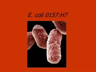
E. coli 3x6K Microarray Slide. Produced by the Microarray and Proteomics Facility in the Department of Biological Sciences at the University of Alberta. Slide Basics. Slide contains Operon’s Version #1 of 3 E. coli genomes Strains represented are K12, O157:H7 (EDL933), and O157:H7 ( Sakai )

E N D
E. coli 3x6K Microarray Slide Produced by the Microarray and Proteomics Facility in the Department of Biological Sciences at the University of Alberta.
Slide Basics • Slide contains Operon’s Version #1 of 3 E. coli genomes • Strains represented are K12, O157:H7 (EDL933), and O157:H7 (Sakai) • 768 controls -12 positive and 12 negative each repeated 32 times • Each spot in triplicate in non-adjacent arrays • Printed on Corning Epoxide Slides for ease of use and low background signal • Printed on GeneMachines OmniGrid 100 with Telechem SMP3 pins
Pargon Dye Test Paragon ss-DNA Binding Dye from Molecular Probes shows presence of DNA on slide with much great resolution and clarity. Actually confirms that DNA is present on the slide. This is a standard part of our QA/QC program.
Paragon Dye –cont- Close up of Mid-group of spots shows excellent detail on spot morphology and spot arrangement after printing. Also shows spots that are buffer controls as defined in the slide map. Example of buffer control, there is not supposed to be DNA here.
Paragon Dye –cont- Close up of middle array from last slide shows benefit of 200 um spot spacing. Spots are very well defined with sizes ranging from 70 um to 110 um with excellent spacing between spots. Spots are also very well formed and concentric as shown in 3D image
3x6K E.coli slide summary • Genomes of 3 strain of E. coli represented in triplicate. • Standard QA/QC test guarantee high quality slide from each print run. • Slides stored/shipped in N2(g) environment in presence of desiccant and O2 scrubber to ensure high quality slides with long shelf life. • QA/QC information, slide maps, slide images are available on-line from our website. • www.biology.ualberta.ca/facilities/microarray